Sunnybrook researchers develop new magnetic resonance imaging methods to help differentiate between aggressive and non-aggressive prostate cancers
Current needle biopsy techniques have limited accuracy in detecting prostate cancer and determining the tumour’s aggressiveness. New methods are needed to better detect and characterize prostate cancer so that each patient can get the treatment that is most appropriate for them.
To address these limitations, researchers at the Sunnybrook Research Institute (SRI) have developed new imaging methods to better characterize and monitor tumours. Their methods, which were recently described in Magnetic Resonance in Medicine, could allow clinicians to identify when and where a prostate tumour becomes aggressive by looking at how cells use energy, or metabolize sugars. This research has received support through OICR’s Imaging Program.
“We know that tumours are made up of many different groups of cells that act, grow, and respond to treatment very differently,” says Dr. Justin Lau, lead of the study and former PhD student in the Cunningham Lab at the SRI. “It’s important for us to get a clear picture of how the tumour is behaving in order to predict how the disease will develop.”
Lau’s recently-published methods use a technique called hyperpolarized carbon-13 imaging, which captures a map of the metabolic activity in the prostate. Clinicians can watch for changes in metabolism as an early sign of a tumour that is starting to become more aggressive and deliver the appropriate level of treatment. For men with prostate cancer, these new methods also offer a more comfortable alternative to traditional biopsies that require 12 needle sticks to collect tissue samples from the prostate.
“We’re developing methods that provide a clearer picture of the cancer to the clinician with less pain to the patient,” says Lau. “This could help patients with aggressive cancers get the treatment that they need, while avoiding unnecessary treatment and the associated negative side effects for those who have latent cancers.”
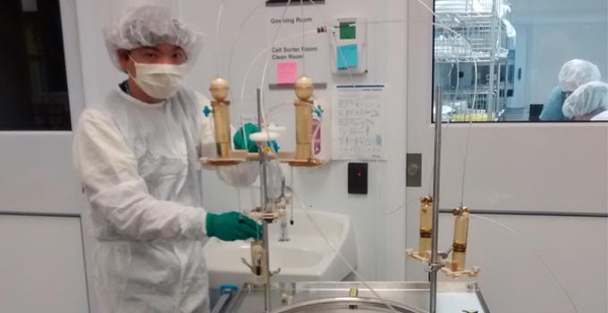
Lau’s imaging methods use GE Healthcare’s SPINlab, the only equipment in the world that can prepare hyperpolarized carbon-13 agents for studies in humans. OICR’s Imaging Program, part of the Adaptive Oncology Platform, has supported the partnership between GE Healthcare and the Cunningham Lab over the past decade. Together, they have improved SPINlab’s technology and associated imaging methods. Like Lau, who is now a postdoctoral research fellow at the University of Oxford, researchers around the world are developing methods that use the SPINlab to study a variety of other cancers and diseases.
“The additional information gained from hyperpolarized carbon-13 imaging could be a real game changer in delivering personalized treatment to prostate cancer patients,” says Lau. “We’re all invested in bringing these methods to the clinic.”