InterConnections
Fortress of Scar Tissue
Receive the latest news, event invites, funding opportunities and more from the Ontario Institute for Cancer Research.
Fortress of Scar Tissue
by Ferris Nowlan and Noor Shakfa, Lunenfeld-Tanenbaum Research Institute
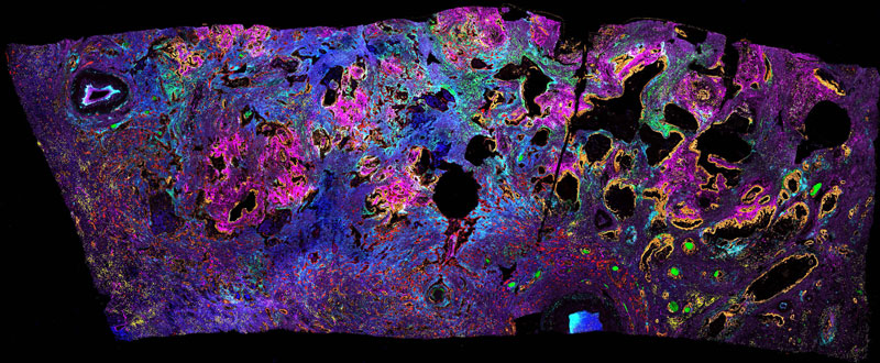
A 1cm x 2cm slice of pancreatic cancer, donated by a patient who underwent surgery. The right half of this image shows one form of the cancer, called the classical subtype (orange), which grows in large ducts. The left and bottom are dominated by a different form of the cancer, called basal subtype (red). The yellow dots are immune cells trying to enter the tumour, but most are kept away from the cancer cells themselves by scar tissue (green, blue, purple, pink). In pancreatic cancer, the scar tissue surrounding the cancer cells can make up as much as 90% of the tumour volume, which is much more than in most other cancers. We are studying how the type of scar tissue changes depending on pancreatic cancer subtype, and how these differences might affect patient response to treatment.
Artist statement
Thank you to all the patients who have donated their tumours to Ontario’s biobanks, and the philanthropists who give to cancer research. I am extremely grateful to be able to work on this project.
Tools and techniques used
Imaging mass cytometry was used to create this image, so the raw data is ion counts per pixel. Colours are computer generated, not natural, as this type of data has no inherent colour. This technique involves staining tissue with antibodies attached to heavy metals with unique atomic masses, and then destroying the tissue one pixel at a time, and measuring the number of ions of each specific weight that were present in each pixel-worth of tissue.
Credits
Ferris Nowlan, PhD Candidate, Lunenfeld-Tanenbaum Research Institute
Noor Shakfa, Postdoctoral Fellow, Lunenfeld-Tanenbaum Research Institute
Hartland Jackson, Principal Investigator, Lunenfeld-Tanenbaum Research Institute

InterConnections is supported by Illumina