InterConnections
Peter Pan Cancer Cells
Receive the latest news, event invites, funding opportunities and more from the Ontario Institute for Cancer Research.
Peter Pan Cancer Cells
by Danielle Harper, Queen’s University
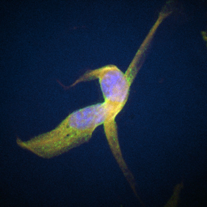
This image features two breast cancer cells, stained for fluorescence microscopy. The nucleus of each cell can be seen in blue, the endoplasmic reticulum in yellow and a cell surface receptor called programmed death ligand-1 (PD-L1) in green. PD-L1 is expressed on the surface of cancer cells to help them evade the immune system. In recent years, new drugs that block the interaction between PD-L1 and its receptor on T-cells have revolutionized cancer immunotherapies.
Artist statement
This image reminds me of the quote: “science is like magic, but peer reviewed”. Like magic, science knows no limits, and provides hope, even in the darkest of times.
Tools and techniques used
Immunofluorescence and confocal microscopy.
Credits
Danielle Harper, PhD Candiate, Sinclair Cancer Research Institute, Queen’s University

InterConnections is supported by Illumina