3D ultrasound prototype shows promise as point-of-care screening tool for women with dense breasts.
Researchers at Western University are hoping their 3D ultrasound system can help close a gap in breast cancer screening for the nearly 40 per cent of women with dense breasts.
Mammography is the gold standard in breast cancer imaging, and regular mammograms can help detect breast cancer early when it is most treatable. But dense breast tissue can ‘mask’ tumours in a mammogram, increasing the chances that early stage cancers are missed.
Ultrasound is a potentially more effective screening option for women with dense breasts. Producing higher contrast images than mammography, standard 2D ultrasound images can help spot cancer independent of breast density. And new developments in 3D ultrasound technology could provide an even more detailed picture.
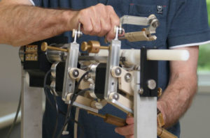
Western University’s Claire Park is first author of a paper in Medical Physics describing a 3D ultrasound prototype for whole-breast imaging. The method involves adding a clinician-controlled robotic arm to an existing 2D ultrasound system, taking 2D photos at different angles, and using custom software to synthesize them into 3D.
“By adding that three-dimensional information, we can better detect cancer, and better characterize the cancer we see,” says Park, a PhD candidate in Medical Biophysics at Western University’s Robarts Research Institute under Dr. Aaron Fenster, who is also Co-Director of OICR’s Imaging Program. “It also opens the possibility for imaging in auxiliary regions of the breast where other systems can’t see.”
Because the prototype is adaptable to any existing 2D ultrasound system — one of the most widely accessible imaging technologies — it has the potential to be rolled out widely and cost-effectively. It also performed well in early testing, producing highly accurate 3D images in volunteers and tissue-mimicking phantoms.
“The results were really encouraging,” Park says. “They show that our system could be used as an accurate alternative [to mammography].”
The study also demonstrated the system’s potential for point-of-care imaging. The prototype is small, fitting onto a medical cart, and Park says it could ultimately be portable enough to screen high-risk women at the bedside.
Though promising, this work is still in early stages. The research team is currently using what it learned from testing to optimize the prototype, working to reduce errors and iterating components to improve workflow. Ultimately, researchers hope to develop a robust protocol for whole-breast imaging and would ultimately like conduct a clinical trial.
For now, Park and her colleagues are encouraged by what their system could mean for a large number of women.
“There is an unmet clinical need for women with dense breasts,” says Park. “A system like ours that harnesses technology that is already readily available in the clinic could really address that need.”
Claire Park was one of six Ontario trainees working in cancer research to receive a 2022 Rising Stars Award.