
Innovative drug screening method finds new promising molecules to treat aggressive lung cancers

Igor and Rima
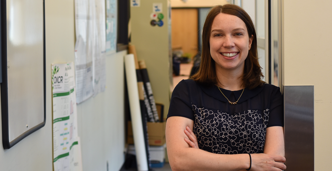
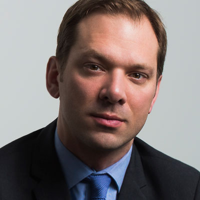
Dr. Igor Stagljar (Photo by Sam Motala) | Dr. Rima Al-awar

International research group finds leukemia drugs and other small molecules may shrink treatment-resistant lung tumours

Lung cancer is the leading cause of cancer death in Canada and around the world. These fatal cancers often arise as a patient’s tumour cells acquire new mutations and become resistant to treatment but Dr. Igor Stagljar has found a new way to stop these tumours. In fact, he may have found four.

Stagljar’s research group at the University of Toronto is well-known for developing a live drug screening method – named MaMTH-DS – that can test potential cancer-fighting molecules in living cells. In a recent study published in Nature Chemical Biology, he and collaborators used these methods to focus on a common mutation, dubbed C797S, which often arises in lung cancers just months after initial treatment. The group identified four new compounds that could block the effects of C797S mutations with no effect on healthy cells.

“Our new technology allows us to find molecules that could be used against cancers for which no other treatment options are available,” says Stagljar, who is a professor of molecular genetics and biochemistry at the University of Toronto. “The advantage of our method is that we are doing it in living cells, where we have all the other molecular machineries present that are important for signal transduction. Also, the compounds are fished at very low dose, which allows us to test for both permeability and toxicity at the same time.”

Conventional drug screening strategies were not able to detect these compounds but Dr. Stagljar’s approach brought these new promising molecules to light.

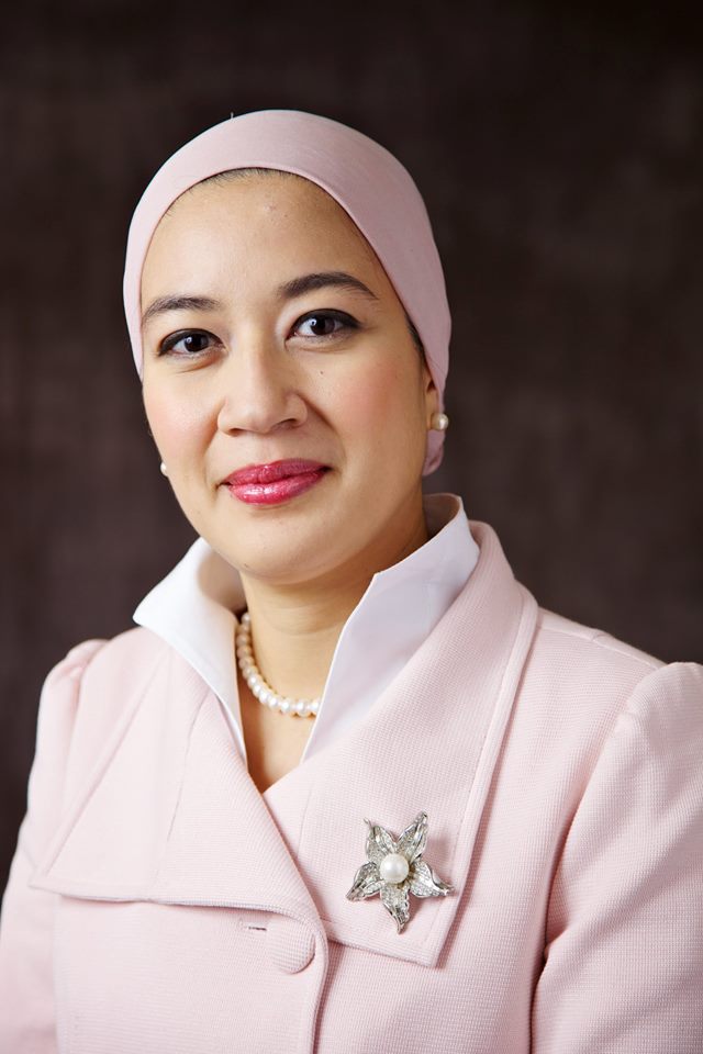
Dr. Rima Al-awar

Two of the molecules identified have already been approved for patients with leukemia. Motivated by their recent findings, Stagljar and collaborators plan to evaluate the effects of these compounds in patients with lung cancer. The first clinical trial to evaluate one of these drugs – gilteritinib – is expected to launch later this year in Toronto, Canada and Zagreb, Croatia.

The other two molecules will require further research and development before they can be trialed in patients. One of these molecules, known as EMI1, could shut down the mutated cells in a completely new way, leveraging molecular machineries to degrade mutated proteins on the surface of tumour cells. The researchers think that EM1’s complex mechanism of action will make it more difficult for tumours to develop resistance to it.

Stagljar is working with Dr. Rima Al-awar, Head of Therapeutic Innovation and Drug Discovery at OICR, and her medicinal chemistry team to create an improved version of the EMI1 molecule. If proven successful, this molecule could potentially become a new treatment for the estimated 60,000 lung cancer patients worldwide who have the C797S mutation.

“Dr. Stagljar’s novel screening approach has identified these very promising molecules” says Al-awar. “We’re proud to collaborate with him and his group to further advance these molecules and accelerate the stages of experimentation between his discovery and helping those with the disease.”

Al-awar, whose drug discovery team recently brought a molecule for blood cancers into pre-clinical development, will leverage her group’s expertise to refine the molecule and move it into the next stage of development, where its ability to shrink tumours can be evaluated in experimental animal models and eventually patients.

This research was supported in part by the Consortium Québécois sur la Découverte du Médicament (CQDM), Cancer Research Society (CRS), Canadian Institute of Health Research (CIHR), Genome Canada and Ontario Research Fund. Stagljar was recently awarded a Prospects Oncology Fund grant from FACIT, OICR’s partner in commercialization, to develop a related drug screening platform, SIMPL.


This post has been adapted from the original announcement made by the University of Toronto Donnelly Centre.
Study shows common cell protein could be targeted to treat childhood brain cancers

Researchers discover that childhood brain cancer could be treated by blocking key cell-surface protein, pointing to a potential treatment approach with fewer toxic side effects

Chemotherapy for children with brain cancer is often toxic, leaving patients with serious life-long side effects but OICR-funded researchers have uncovered a new approach that may help.

In a study published in the Journal of Experimental Medicine, the Ontario-based research team discovered that blocking a specific protein on the surface of brain cancer cells can suppress the rampant growth of a tumour without harming the development of the brain.

The study focused on the protein CLIC1 in medulloblastoma, the most common type of childhood brain cancer. The group found that disrupting CLIC1 can halt medulloblastoma growth with very little effect on the developing brain in mice.

“Brain cancer is the leading cause of cancer-related death in children and young adults,” says Dr. Xi Huang, Scientist in the Developmental & Stem Cell Biology Program at The Hospital for Sick Children (SickKids) and senior author of the study. “We need new treatments to help these patients.”

We believe our findings are significant because ion channels have been successfully targeted to treat numerous human diseases.

Michelle Francisco

CLIC1 belongs to a class of proteins called ion channels, which are important in the development of several other diseases like diabetes, epilepsy and high blood pressure. Many existing drugs and compounds act as ion channel modulators. The Huang Lab now has the high-throughput screening equipment to assess thousands of drug-like chemicals for those that can best block these ion channels.

“We believe our findings are significant because ion channels have been successfully targeted to treat numerous human diseases,” says Michelle Francisco, Research Project Coordinator in the Developmental & Stem Cell Biology Program at SickKids and first author of the study. “This helps pave the way between this discovery today and the impact it can have in the clinic.”

These findings build on Huang’s previous research on the potassium channel EAG2, which – like CLIC1 – is critical to medulloblastoma growth. In partnership with collaborators, Huang has shown that EAG2 could be blocked with an FDA-approved drug for schizophrenia to treat medulloblastoma in experimental mouse models and in a small patient study.

“We are fortunate to work with world-leading brain cancer researchers in Ontario,” Huang says, “We look forward to continuing our research to find new solutions for this devastating disease by targeting ion channels.”

This research was funded by OICR’s Brain Cancer Translational Research Initiative, SickKids Foundation, Arthur and Sonia Labatt Brain Tumour Research Centre, Garron Family Cancer Centre, b.r.a.i.n.child, Meagan’s Walk, Natural Sciences and Engineering Research Council (NSERC) Discovery Grant, U.S. Department of Defense (DoD) Peer Reviewed Cancer Research Program Career Development Award, Canadian Institute of Health Research (CIHR) Project Grants, and Sontag Foundation Distinguished Scientist Award to Xi Huang.

Read more about OICR’s brain cancer research.

Harnessing the time before a patient’s surgery to accelerate cancer research

Ottawa cancer researchers and clinicians embrace the window of opportunity between a cancer diagnosis and treatment with a coordinated approach to clinical research

The time between a patient’s cancer diagnosis and their surgery presents a valuable “window of opportunity” to evaluate new treatment strategies. Short-term clinical trials during this period – also known as window of opportunity trials, window trials or phase 0 trials – can help researchers gain insights into the effects and the efficacy of a new potential treatment. Dr. Angel Arnaout at The Ottawa Hospital is putting window trials into practice.

“There are many nervous and anxious moments between diagnosis and their surgery but patients have limited options during this time,” says Arnaout.

“We saw an opportunity in this window of time to take action. We saw that we could help support patients who are waiting for surgery, while helping future patients through accelerating clinical research.”

Dr. Angel Arnaout

Arnaout, a surgical oncologist who specializes in breast cancers, assembled a cross-disciplinary team of medical oncologists, pathologists and other clinical research specialists at The Ottawa Hospital to strategically design and implement this new approach. They would collectively establish common priorities, decide on which interventions would be tested and work to streamline the patient’s journey throughout the process.

Dr. Angel Arnaout

Together, the team was motivated by the mutual benefits of all stakeholders involved. Namely, window trials can provide patients an opportunity to contribute and engage with cancer research while potentially improving the state of a patient’s disease. Meanwhile, these trials could ultimately expedite drug development by improving the understanding of a potential drug early in its development.

The team launched their first study in 2014, which found that patients were exceptionally eager to participate, and since then, launched and completed three additional window trials.

The first was a breast cancer trial on presurgical hormone therapy that helped establish the capacity and infrastructure for enrolling patients, organizing the investigations and giving patients short-term therapies. The second tested a potential cancer-fighting agent, chloroquine, and found that it had no effect on stopping breast cancer proliferation. The third trial debunked the idea that vitamin D – even at very high doses – can slow down the growth of breast cancer.

“These studies didn’t uncover a new therapy, but they did help us answer important questions that patients have, like ‘Will taking vitamin D help?’” says Arnaout. “These types of studies also provide a relatively quick method to test whether we should continue research into a particular avenue.”

The group at The Ottawa Hospital has recently teamed up with researchers from OICR to initiate a new breast cancer window-of-opportunity study to examine biomarkers of efficacy and resistance for another new drug candidate. The trial is planned to begin recruitment by mid-fall this year.*

Despite the benefits of these trials, Arnaout adds, it is still important to reduce unnecessary delays between diagnosis and surgery. Arnaout continues to minimize these delays at The Ottawa Hospital.

“We try our best to reduce wait times, but if patients have to wait – we can try to help them in the meantime while accelerating breast cancer research.”

*This new trial is co-led by Dr. John Hilton from The Ottawa Hospital and Dr. John Bartlett from OICR. Co-investigators include Drs. Laszlo Radvanyi, Melanie Spears, Arif Ali Awan, Mark Clemons, Greg Pond and Angel Arnaout.

Meet, network, connect at the Canadian Cancer Research Conference

The Canadian Cancer Research Association releases their calendar of special events for attendees to meet, connect and network with peers at the Canadian Cancer Research Conference.

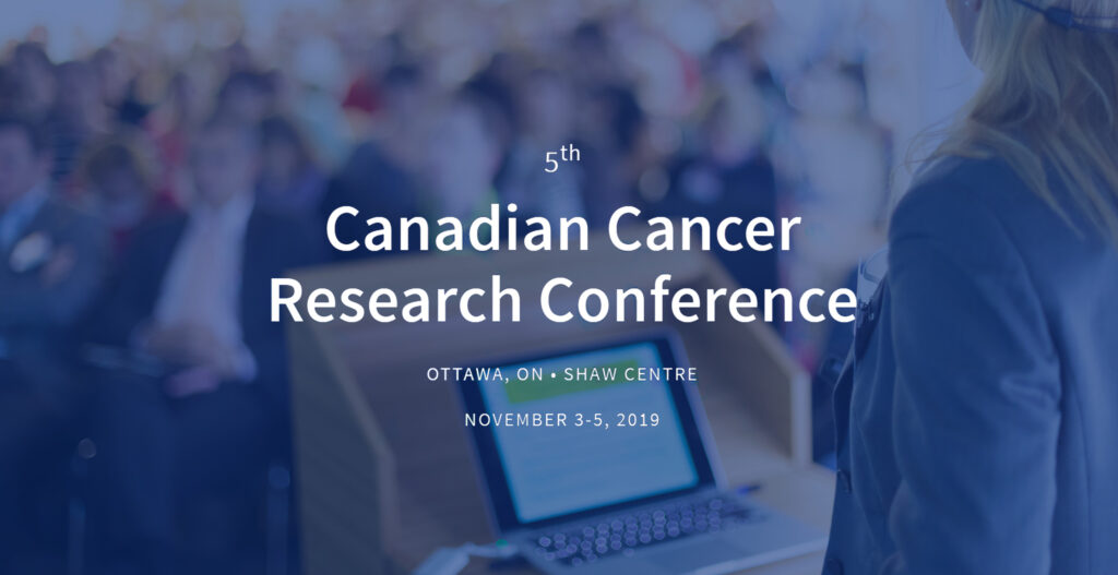
5th Canadian Cancer Research Conference, Ottawa, ON, Shaw Centre, November 3-5, 2019

The 5th Annual Canadian Cancer Research Conference, which will be held November 3-5 in Ottawa, will bring together cancer researchers across disciplines, interests, and at all stages of their career to explore the latest advancements and knowledge in the field.

Explore the special events below or read more on the event website.


All-ages community event – Cancer: Piecing the Puzzle Together
Canadian Cancer Research Alliance 
Saturday, November 2, 2019 
12:30 pm -4:30 pm EST

Everyone is welcome to attend this free, family-friendly and bilingual event which will feature English and French sessions. The event will display a giant, inflatable model of a human colon, have an impressive lineup of speakers, and showcase the many ways cancer research is saving lives and revolutionizing patient care.


(more…)

Computational dissection: What can we learn from the cells around cancer cells?

OICR researcher looks into what non-tumour cells can tell us about breast cancer

Natalie Fox
Natalie Fox, OICR

When a biopsy is drawn from a patient, it consists of a mix of cancerous and healthy cells, like fat and blood cells. Researchers are often interested in diseased cells, but without looking into the surrounding tissue, they could be missing part of the story.

Natalie Fox, a PhD candidate at OICR, is investigating what we can learn from the cells surrounding cancer cells.

“When we look into a patient sample computationally, we see distorted signals because of overlapping data from many different types of cells,” says Fox. “We need to dissect the parts we want to study, but instead of using a knife or a laser, we use computers.”

Fox and collaborators have analyzed nearly 1800 tumour samples from patients with breast cancer, examining the transcriptome of tumour cells and the cells around tumours – or the tumour’s microenvironment.

Her study, recently published in Nature Communications, reveals the landscape of transcriptomic interactions between breast cancers and their microenvironments. Her study also sheds light on associations between these transcriptomes and patient survival, gene mutations and breast cancer subtypes.

“We now have a clearer picture that tells us more about the breast microenvironment than we’ve known before,” Fox says. “Bit by bit, we’ve analyzed and scrutinized these data, then assembled these bits into a comprehensive landscape.”

Fox found that mutations in cancer genes such as CDH1 and TP53 are associated with changes in the transcriptome of the tumour’s microenvironment. She says more research is needed to clarify the biologic rationale behind her observations, but her work has set the stage for researchers to do so.

“Above all else, this work demonstrates an important approach for improving our understanding of associations between the tumour and the microenvironment,” Fox says. “We presented a framework that others can use to analyze the tumour microenvironment in their cancer of interest and potentially develop new biomarkers for predicting cancer patient outcomes.”

Blood samples, biostatistics and a fresh perspective: The makings of a cancer prediction machine

Biostatistics Training Initiative (BTI) alumnus brings on new BTI trainee to study Canada’s largest population health dataset using today’s top technologies

Recently, circulating tumour DNA (ctDNA) – DNA released from cancer cells that freely circulates in the blood – has garnered much attention not only as an alternative to traditional tissue biopsies, but as a potential blood-based biomarker for early cancer diagnosis.

The ability to detect the earliest blood-borne traces of cancer largely rests in our ability to determine which molecular markers indicate that a cancer is developing – or which patterns in ctDNA can predict whether a cancer will grow. Dr. David Soave sees this as a mathematical challenge that, if solved, could have huge impact for better predicting and diagnosing a wide variety of cancers.

“To find cancer earlier or predict who will develop the disease, we need to carefully compare human samples from those who will develop cancer and samples from those who won’t,” Soave, an Assistant Professor at Wilfrid Laurier University and OICR Associate, says. “This type of challenge requires new statistical models, methods and computational techniques that can decipher large, complex and high-dimensional data.”

Last year, the Canadian Partnership for Tomorrow Project (CPTP) unified the data from several provincial longitudinal health studies into a national cohort consisting of more than 325,000 participants who are voluntarily donating their health and biologic samples to research. As some CPTP participants will develop disease and others will not, this dataset provides an unprecedented resource for researchers like Soave to discover the earliest traces of cancer that appear several months to years prior to an initial diagnosis.

(more…)

New research unfolds the DNA “origami” behind brain cancers

Collaborative research group maps the three-dimensional genomic structure of glioblastoma and discovers a new therapeutic strategy to eliminate cells at the roots of these brain tumours

Current treatment for glioblastoma – the most common type of malignant brain cancer in adults – is often palliative, but new research approaches have pointed to new promising therapeutic strategies.

A collaborative study, recently published in Genome Research, has mapped the three-dimensional configuration of the genome in glioblastoma and discovered a new way to target glioblastoma stem cells – the self-renewing cells that are thought to be the root cause of tumour recurrence.

The research group integrated three-dimensional genome maps of glioblastoma with other chromatin and transcriptional datasets to describe the mechanisms regulating gene expression and detail the mechanisms that are specific to glioblastoma stem cells. They are one of the first groups in the world to perform three-dimensional genomic analyses in patient-derived tumour samples.

Dr. Mathieu Lupien

“The 3D configuration of the genome has garnered much attention over the last decade as a complex, dynamic and crucial feature of gene regulation,” says Dr. Mathieu Lupien, Senior Scientist at the Princess Margaret Cancer Centre, OICR Investigator and co-author of the study. “Looking at how the genome is folded and sets contacts between regions tens to thousands of kilobases apart allowed us to find a new way to potentially tackle glioblastoma.”

Through their study, the group discovered that CD276 – a gene which is normally involved with repressing immune responses – has a very important role in maintaining stem-cell-like properties in glioblastoma stem cells. Further, they showed that targeting CD276 may be an effective new strategy to kill cancer stem cells in these tumours.

Lupien adds that advancements in three-dimensional genomics can only be made through collaborative efforts, like this initiative, which was enabled by OICR through Stand Up 2 Cancer Canada Cancer Stem Cell Dream Team, OICR’s Brain Cancer Translational Research Initiative and other funding initiatives.

“This research was fueled by an impressive community of scientists in the area who are committed to finding new solutions for patients with brain cancer,” Lupien says. “Our findings have emphasized the significance of three-dimensional architectures in genomic studies and the need to further develop related methodologies to make sense of this intricacies.”

Senior author of the study, Dr. Marco Gallo will continue to investigate CD276 as a potential therapeutic target for glioblastoma. He plans to further delineate the architecture of these cancer stem cells to identify more new strategies to tackle brain tumours.

Dr. Marco Gallo

“A key problem with current glioblastoma treatments is that they mostly kill proliferating cells, whereas we know that glioblastoma stem cells are slow-cycling, or dormant. Markers like CD276 can potentially be targeted with immunotherapy approaches, which could be an effective way of killing cancer stem cells, irrespective of how slowly they proliferate,” says Gallo, who is an Assistant Professor at the University of Calgary. “Being able to kill cancer stem cells in glioblastoma could have strong implications for our ability to prevent relapses.”

Read more about OICR’s Brain Cancer Translational Research Initiative on oicr.on.ca or read about the Initiative’s current findings on OICR News.

Understanding intermediate-risk prostate cancer: The whole may be greater than the sum of its parts

Collaborative research group performs the most comprehensive analysis of curable prostate cancer to date, finds key connections between different data types

As cancer researchers delve deeper into different omics studies, and technologies enable their ability to do so, it is becoming increasingly important to understand how these areas of research are interconnected. Previous studies across multiple omes – such as the genome, proteome, transcriptome or epigenome – have led to important discoveries in colorectal cancer and ovarian cancer, but prostate cancer remains largely unresolved. Researchers from the Canadian Prostate Cancer Genome Network (CPC-GENE) set out to unravel some of these mysteries.

In the most recent CPC-GENE study, published today in Cancer Cell, the research group integrated multiple levels of omics analyses to better understand the biology of intermediate-risk prostate cancer – a type of cancer in which it is notoriously difficult to predict and treat accordingly. A better understanding of this disease could lead to improved tests that can determine which tumours are aggressive and require aggressive treatment, while helping spare those whose cancer will never become aggressive the negative side effects of treatment.

“We cannot overlook the important information that we gain from looking at the bigger picture,” says Julie Livingstone, bioinformatician at OICR and co-author of the study. “In this case, this means looking at prostate cancer from multiple angles – or multiple omes – to potentially find new markers of aggressive disease.”

The study explored 76 prostate cancer tumours and found new combinations of information that could act as a better predictor of a patient’s chance of relapse than any single piece of information alone. More specifically, they identified that the combination of protein and methylation data could, on average, predict the severity of a tumour better than looking at just the proteins – the proteome – or just the methylation patterns – the methylome – alone.

“Integrating datatypes is anything but straightforward, but it illuminates interesting aspects about prostate cancer that we haven’t seen before,” says Livingstone. “In the future, we intend to pursue our multi-omic investigation and translate this understanding into better tools to inform treatment selection for men with this disease.” 

Find out more about research from the CPC-GENE project on OICR News.

Towards rational breast cancer treatment in the era of “less is more”

Expert researchers find shorter treatment cycles may reduce risk of breast cancer returning

Researchers have found that the dosage and interval of chemotherapy treatments have a significant impact on some breast cancer patients’ survival. For a very small minority of patients the difference of a week between chemotherapy treatments could mean the difference between life and death – and researchers are working to identify exactly who those patients are.  

Over the last few decades, breast cancer clinical trials have investigated the way in which patients receive and respond to different chemotherapy dosing regimens. Some have tested if a shorter – but more intense – two-week treatment cycle is more effective than the standard three-week cycle. These trials, however, are often limited in size and do not have the statistical power to detect a difference in response to treatment.

Researchers from the Early Breast Cancer Trialists’ Collaborative Group (EBCTCG) have recently performed a meta-analysis across 26 breast cancer trials to help clarify this dosing dilemma. As reported in The Lancet, they found that more intense dosing regimens were associated with a decreased risk of death from breast cancer and a decreased risk that the disease will return in some patients.

“As chemotherapy kills tumour cells, the residual – or remaining – cancer cells have more room to grow and tend to grow faster,” says Dr. John Bartlett, Program Director of Diagnostic Development at OICR and member of the EBCTCG Steering Committee. “These trials hypothesized that a more intense dosing regimen would give tumour cells essentially less room to grow. With the results of the EBCTCG overview study, we can say with confidence that a two-week treatment schedule will help to prevent death in a small portion of patients.”

The analysis found that approximately one in 50 women benefited from more intense dosing.

“The challenge now is to determine exactly which patients can benefit from intense dosing and which patients would not,” says Bartlett.

“If we can do so, we can prevent deaths due to breast cancer for some, while minimizing the negative side effects of intense chemotherapy for others.”

The ECBTCG is continuing to investigate dosing intensity in common breast cancer subtypes in parallel with researchers who are looking to find the biological basis of these differing responses to treatment.

“We’re in an era of de-escalation where we’re heavily invested in reducing overtreatment,” Bartlett says. “But this work helps us move towards an era of biologically rational treatment recommendations, one where breast cancer patients get the treatment they need at the right time and in the right way.”

When it comes to prostate cancer, there may be more than meets the eye

Researchers begin to unravel why some prostate tumours can be seen with magnetic resonance imaging and others go undetected

Determining whether a patient with prostate cancer requires aggressive therapy or active surveillance is a growing challenge for the healthcare system. Blood tests can detect early signs of prostate cancer, but these tests can lead to many unnecessary and painful biopsies for patients whose disease never becomes aggressive.

Multi-parametric magnetic resonance imaging (mpMRI), a type of non-invasive imaging technique, has the potential to help determine which patients require biopsies and which can be spared possible negative side effects, such as bleeding, pain and infection. Some tumours are visible by mpMRI while some are not, yet it’s not well understood if this visibility can predict a tumour’s aggressiveness.

Researchers at OICR have teamed up with clinicians from the University of California, Los Angeles to investigate the molecular properties of MRI-visible and MRI-invisible tumours. In their recent study, published in European Urology, they found that visible tumours have similar features to aggressive tumours and discovered new features that may be contributing to the disease’s aggression. 

“Even if two tumours are similar in size and in similar positions, one still may be MRI-visible and one may be MRI-invisible,” says Kathleen Houlahan, PhD Candidate at OICR and lead author of the study. “We wanted to see if this visibility could help us determine if a cancer is aggressive, so we took the first step towards unraveling the relationship between a patient’s MRI results and the molecular characteristics of their tumour.”

Recent commentary on the study highlights Houlahan’s work as an “initial foray” into the intersection of radiology, pathology and genomics, but recognizes the limited size of her exploratory study. Recent MRI-focused clinical trials will provide larger datasets for further investigation.

 “If we can better understand why some tumours show and some don’t, we could potentially use imaging to predict the course that a patient’s disease will take,” says Houlahan. “Ultimately, we hope that this technique can help reduce unnecessary prostate biopsies and ensure that the men who need treatment get the treatment they need.”