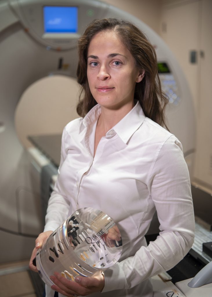
Dr. Catherine Coolens with new DCE-CT calibration device
OICR offers new CT calibration service as part of its Collaborative Research Resources portfolio
Using imaging devices to help make treatment decisions in the clinic requires rigorous testing, quality assurance and routine calibration of the imaging machinery. These standards are especially important when the imaging technology is novel or unique, such as in the case of perfusion imaging – a relatively new technique used to diagnose a cancer’s stage by showing how blood flows through the tumour.
OICR-funded researchers have found a new way to easily perform quality assurance testing on dynamic contrast-enhanced computed tomography (DCE-CT) scanners – today’s cutting-edge perfusion imaging technology. They’ve specially designed a new instrument, which is now used in clinical trials, that can tune the performance of these scanners so that clinicians can better determine the stage of their patient’s cancer and monitor response to treatment.
“Perfusion imaging has the potential to help oncologists determine the best treatment for their patient’s cancer and tailor treatments accordingly,” says Dr. Catherine Coolens, Scientist at the Princess Margaret Cancer Centre. “Calibrating and standardizing DCE-CT devices is necessary so that we can use this technology to its full potential.”
Their instrument, which was recently described in the European Journal of Radiology, consists of an acrylic cylinder with markings and capsules that act as guides to measure the performance of the DCE-CT scanner. The instrument incorporates multiple imaging parameters into one simple device so that the scanner can be calibrated easily, with better accuracy than ever before.
“Before we began, there was a complete lack of validation that these CTs could be used reliably,” says Coolens. “Now that we’ve developed the standards and the instruments necessary to assess DCE-CT scanners, we can use these technologies with more confidence.”
Coolens’ instrument and quality assurance methods were developed at the Princess Margaret Cancer Centre and are now used at multiple cancer centres in Toronto and London, Ontario. Her methods are offered to Ontario’s research community through QIPCM, an OICR Collaborative Research Resources program that provides imaging services and tools to enable multi-centre clinical trials across the province.
Researchers who are interested in accessing these services should contact CRR@oicr.on.ca.

