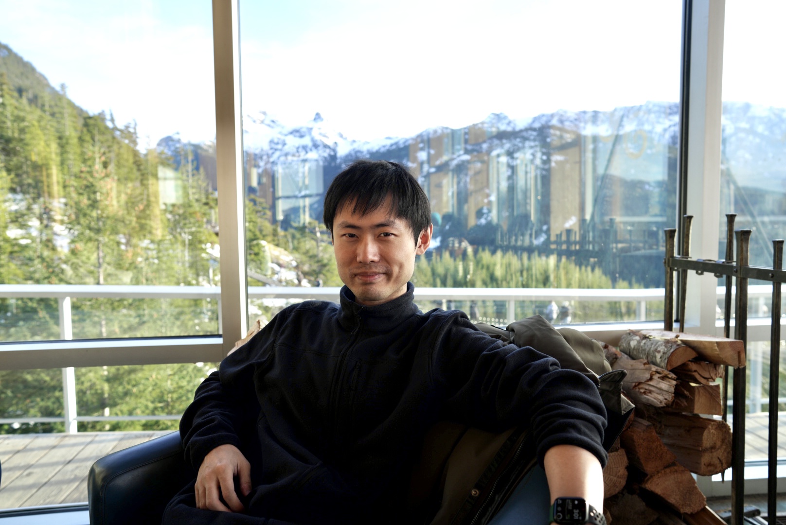
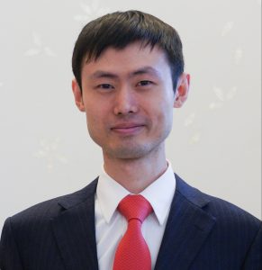
Dr. Chenxi Qian has developed an advanced vibrational spectroscopy technique that delivers fast, high-resolution and label-free imaging.
Dr. Chenxi Qian is excited to take advanced concepts from physics and chemistry and apply them to real-life problems.
Qian is a physicist and chemist who specializes in vibrational spectroscopy, a method for measuring the chemical bonds of different molecules. Though one type of vibrational spectroscopy – known as ‘infrared’ – has some well-known applications in health science, Qian’s specialty – known as ‘Raman spectroscopy’ – isn’t well-known by your average clinician.
But Qian hopes to change that. The Raman method he developed has the potential to be a faster and more accurate tool to scan tissue samples for diseases like cancer.
“In healthcare, patients can’t afford to wait a long time for scans to tell them a diagnosis or prognosis,” Qian says. “We want to bring clinicians a new tool to help them deliver personalized medicine.”
Originally from China, Qian did his undergraduate studies Nanjing University before moving to Canada to earn his PhD in chemistry from the University of Toronto. Qian’s interest in spectroscopy led him south of the border to the California Institute of Technology (Caltech), where he did his postdoctoral fellowship. Now, he has come back to Canada to become Assistant Professor of Medical Biophysics at the University of Toronto and to start his own lab at Sunnybrook Hospital.
Earlier this year, Qian received an OICR Early Career Investigator Award to support research into his innovative imaging method. OICR News recently asked him about his method, and about what drives him as a scientist.
When did you know that you wanted to become a scientist?
I was interested in science from a very young age. When I was in elementary school, we would sometimes give speeches to the class. My speeches were usually about physics or chemistry – I would learn some advanced, abstract concept and then teach it to my classmates. Then, in high school, I decided I wanted to become a scientist and a professor.
Were you also interested in healthcare early on?
That interest didn’t come until later. My graduate studies were in physics and chemistry. Then during my postdoctoral fellowship at Caltech, I focused mainly on a method based on nonlinear vibrational spectroscopy called ‘stimulated Raman scattering imaging’.
Raman microscopy is an imaging technique that has been used for many years in different fields, but I believe its future lies in healthcare. With recent advances, it can help create a 3D image of the components of a particular sample, and that can be very useful in healthcare, and especially in cancer.
Also, like many people, I have a personal connection to cancer. When I was very young, I lost my grandfather to lung cancer. He was very dear to me. I believe this was a signal to me that I should use my knowledge to contribute to cancer research.
What exactly is vibrational spectroscopy and what makes it effective for imaging in healthcare?
Vibrational spectroscopy is a way to visualize the molecules in a sample by probing their molecular vibration. Each chemical bond within molecules has a specific vibrational frequency, whether the molecules are proteins or lipids or something else.
The advantage of this imaging technique in healthcare is that it is label-free. The current standard for tissue samples is histology, where dyes are used to stain the sample. Sometimes that staining process is very long and troublesome and doesn’t allow for quantification. The Raman imaging method we use can achieve very high resolution without the labeling process and all that comes with it.
You have developed an innovative technique for Raman imaging in healthcare. Can you tell us more about that?
In the lab I’m building at Sunnybrook, we use an advanced form of Raman imaging called ‘simulated Raman scattering imaging’. I contributed to advancing this technique at Caltech with my colleagues and supervisors. We created a way to enhance the Raman signal to achieve high-resolution, high-throughput imaging. We are also combining this with machine learning methods, so that we can get an even faster throughput.
We are currently building our machine. When we’re done, we want to bring it to clinicians so they can have faster, label-free imaging to support personalized medicine.
What excites you about the potential of this technique?
A lot of physicists and chemists come up with a method, and then leave it to others to worry about its application. But I am taking the opportunity to work on the application of our method myself, because I believe it can be very valuable in healthcare. I’ve also had the opportunity to talk to many clinicians about my method. They also think it could be useful to them, and that’s very exciting to me.


