Prostate cancer trial aims to deliver shorter, more effective radiotherapy thanks to advanced imaging

Sophisticated imaging tools are being used together to guide high-dose radiation that provides safe and effective treatments in a shorter timeframe.


For John Hodges, the math was simple. Five radiation treatments sounded better than 28.

The 74-year-old Goderich resident was diagnosed with prostate cancer earlier this year and told he wasn’t eligible for surgery because of pre-existing health conditions.

His doctors recommended radiation therapy. But that would mean driving three hours round trip from Goderich to the nearest cancer centre in London for 28 treatments over the course of several weeks.

“I don’t know if I could do 28 treatments in a row, driving back and forth. It would be awful,” Hodges said.

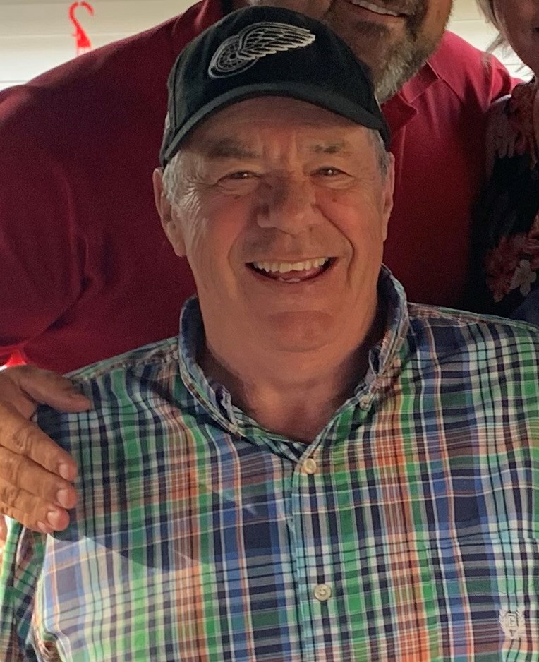
John Hodges, the first participant in the ARGOS/CLIMBER clinical trial.

Then his doctors told him about an innovative clinical trial where he could safely get higher doses of radiation and would only need five treatments over two weeks.

“It made a lot of sense to me,” he says. “I asked a lot of questions, the doctors answered every one of them, and I decided to go ahead with it.”

Hodges was the first participant in ARGOS/CLIMBER, a newly launched clinical trial led by Dr. Glenn Bauman of Lawson Health Research Institute and London Health Sciences Centre and Dr. Andrew Loblaw of Sunnybrook Hospital.

The first part of the trial focuses on combining two innovative imaging techniques — prostate-specific membrane antigen positron emission tomography (PSMA-PET) and multiparametric MRI (mpMRNI) — to guide a high-dose radiation procedure called stereotactic body radiation therapy (SBRT).

PET scanning is a sophisticated technique where a molecule designed to seek out and bind to cancer cells is injected into the body to highlight the presence of cancer. mpMRI is a prostate-specific scan that can provide a more detailed image than a standard MRI. Together, they can pinpoint the location of the prostate and find the ‘dominant’ area with the highest concentration of cancer cells.

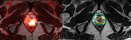
Example of a PSMA PET/MRI demonstrating a focus of tumour in the prostate gland (left image) and a radiation plan boosting the tumour to an escalated dose compared to the remainder of the prostate (right image).

Localizing the prostate and the cancer within it is crucial to SBRT, which involves delivering a concentrated dose of radiation to a precise target. The highly detailed images from PSMA-PET and mpMRI add an extra level of precision to the process, eliminating the need for the large safety margins used to protect the surrounding organs from radiation.

“If we know where the cancer is within a millimeter or two then we can shrink those safety margins and safely increase the dose of radiation to that dominant area within the prostate,” says Bauman.

Techniques like PSMA-PET, mpMRI and SBRT are the latest steps in a series of improvements to radiation therapy that have been driven by advances in imaging. Ten years ago, when radiation treatment was mainly guide by CT scanning, it took up to 40 doses of radiation over several weeks to treat prostate cancer.

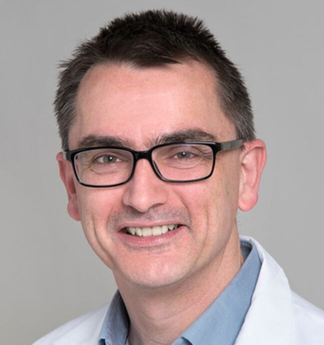
Dr. Glenn Bauman

“These advances allow us to deliver a more effective dose in a way that is more convenient for the patient, and also more resource efficient,” Bauman says. “That’s a win-win for everyone.”

Confirming that radiation was effective is the second part of the ARGOS/CLIMBER trial. Participants will have PSMA-PET and mpMRI scans six months and two years after their treatment to assess how their cancer responded and whether it is likely to recur.

These post-treatment assessments are usually done with a prostate-specific antigen (PSA) test or by taking a biopsy of the prostate. But in both cases, it can be hard to confidently know that cancer is gone until at least a few years after treatment.

“It creates a lot of uncertainty for men during that time,” Bauman says.

Bauman and colleagues hope that the combination of PSMA-PET and mpMRI can accurately assess a patient’s response to treatment and find recurrence early, when additional treatment is most effective. By analyzing these sophisticated scans before and after treatment, they also hope they can find clues about how different types of cancers respond to radiotherapy.

“One goal of this study is to better understand how these scans evolve over time after treatment,” Bauman says. “Just by working with scans it’s quite remarkable what you start to see.”

The ARGOS/CLIMBER study is supported through the Clinical Translation Pathway (CTP), a program within OICR’s Clinical Translation initiative, which is designed to accelerate the translation of cancer research into treatments that benefit cancer patients.

“The CTP program supports practice-changing research in biomarkers, diagnostics and therapeutics that will advance early detection and intervention research and have a clear path to the clinic and clinical impact,” says Dr. Steven Gallinger, Lead, Clinical Translation. “Given that prostate cancer is a significant public health concern, improving primary radiotherapy particularly after the detection of early recurrence would be of tremendous value. The promise of personalizing treatment from the ARGOS/CLIMER study is exciting.”  

Hodges is still months away from his six-month scan. But a few weeks after his last radiation treatment, after some gastrointestinal side effects passed, he was feeling good.

He says his five days of extensive scanning and radiation were long and tiring but he’s grateful for Dr. Bauman and his staff and he appreciates his compressed treatment schedule. Now, he’s hopeful that his treatment was successful.

“I feel like I caught the cancer in time, and now I’m putting my faith in the doctors,” Hodges says.

Trusting the gut in cancer research: Q&A with Dr. Saman Maleki

OICR’s new Early Career Investigator is harnessing the gut microbiome’s immunogenic properties to improve cancer patients’ response to immunotherapy.

To make progress against a disease as complex as cancer, Dr. Saman Maleki thinks it’s important to consider unconventional approaches. His groundbreaking research combines his backgrounds in immunology and microbiology with a passion for translational research that makes a difference for people with cancer.

Maleki’s work at Western University and the Lawson Health Research Institute focuses on novel approaches to sensitize tumours to immunotherapy, and he helped pioneer the use of fecal microbiota transplantation (FMT) to strengthen cancer patients’ immune systems.

Now, as OICR’s newest Early Career Investigator, he aims to move his research forward to open up new therapeutic options against other hard-to-treat cancers such as pancreatic cancer.

OICR News recently spoke to Maleki about his research, his motivation and his hopes for the future of oncology.


Tell us about your research interests and your approach to your work.

I’ve always been interested in translational cancer research and what happens to our research after it leaves the lab. As scientists, we want our work to have an impact on patients. But I’ve learned that doesn’t happen easily.

As a PhD scientist, I was determined to help bridge the gap between basic science and the clinic. So, I spent a lot of time with medical oncologists and other clinicians, speaking to them and going to see patients with them. That helped expose me to what happens in the clinic, and I started identifying areas where basic science could help address some of their concerns. The time I spent in the clinic also reinforced the importance of coming up with innovations that can open new lines of treatment for patients.

Immunotherapy is an exciting new class of cancer treatment. Tell us about your research into the microbiome and what it could mean for immunotherapy.

The gut microbiome is the bacteria and other bugs that live in your gut, and research has shown there is a very close relationship between the gut microbiome and the development of the immune system. The two systems are always interacting, both in health and in disease.

Looking at the literature, it seemed that when certain features in the microbiome were present in a patient, they tended to respond better to immunotherapy drugs called checkpoint inhibitors.

The beauty of the microbiome is that is a malleable organ. You can change the microbiome through procedures like FMT, where stool and the bacteria within it are transplanted from one person to another. So, I thought: why don’t we try using FMT to change cancer patients’ microbiome before they have immunotherapy and see if we can improve their response?

You were among the first researchers to propose using FMT in cancer patients. What was the reaction from the oncology community?

We started designing our first trial back in 2016, and there was not much interest in the microbiome back then. There were hints in the literature that the microbiome could be used as a therapeutic modulation strategy for cancer but I’m not sure the field was ready for it at the time. It was considered very ‘outside the box’ and I even had oncologists walk away from me when I was explaining the idea.

But I was convinced that we could replace a cancer patient’s microbiome with a healthy microbiome and potentially improve their response to immunotherapy. And there were some brave oncologists who went over the literature and the theory behind it with me and became interested. After a huge amount of effort by many people, we gradually built a program of prospective clinical trials where we performed FMT in cancer patients before they had immunotherapies.

Through these trials, we’re now providing a new treatment in cancer patients that wasn’t there before, and not a day goes by where I don’t get a call from someone interested in our work.

What is the status of these trials and what have you found so far?

Our first trial was in advanced melanoma patients at London Health Sciences Center in London and two other hospitals in Montreal (CHUM and JGH), and it was recently featured in an article in Nature. Of the 20 people we enrolled, 65 per cent had either a partial or complete response to immunotherapy after having FMT. Though it is preliminary, the data we collected was remarkable and we are very encouraged by the safety signal.

Using what we learned from that melanoma trial, we built other trials using FMT before immunotherapy in renal and lung cancer patients, and we are just starting the process of setting up a trial in pancreatic cancer.

What are the biggest questions remaining about the microbiome and immunotherapy?

I think we still have a lot to learn about the functionality of the microbiome, and about the ideal composition of the microbiome to improve response to immunotherapy. That’s tricky because everyone’s microbiome is different, and the ideal composition for a melanoma patient might be different than for a lung cancer patient.

We also need to determine the best approaches to target the microbiome. The transplant we are doing is sort of the ‘nuclear option’ where you get the entire gut microbiome from someone and put it in a new person. But there could be more targeted options that better meet an individual patient’s needs. More research is required to understand which patient would need a more robust change in their gut microbiome or a more targeted approach.

Where do you see this work going in the future?

Oncology is shifting and we are entering a new era where we are focused more on treating the patient instead of treating their tumour.

Immunotherapies will help define this new era, but at the moment, a large proportion of people are resistant to them. Our work is a proof of principle that you can safely target the microbiome with the goal of improving immunity and ultimately increasing response to immunotherapies.

With the knowledge we’ve gained, I can foresee a future where we profile a patient’s microbiome for deficiencies the same way we profile mutations in cancer looking for druggable mutations. Then, based on the signatures we find, we can build microbiome approaches on top of the other available treatment options.

That is truly personalized medicine, and it gives us one more approach to bring to the table against cancer.


Funding from Ontario Institute for Cancer Research to help move research from experiment to impact

First round of Innovation to Implementation Supplement (I2IS) grants awarded to five promising research projects taking unique approaches to addressing cancer.

August 11, 2022, TORONTO – Five Ontario-based research projects will take the next step toward advancing cancer care in the province thanks to funding from the Ontario Institute for Cancer Research (OICR).

OICR is announcing the first round of recipients of its new Innovation to Implementation Supplement (I2IS), which aims to help discoveries in cancer overcome barriers to health system implementation. All five projects advance research programs that were previously funded by OICR within the past five years. The supplemental funding announced today completes the continuum for these projects, helping them build on their previous success and translate what they have already learned into real-world impact.

“With Ontario’s population aging and the number of cancer diagnoses on the rise, translational cancer research that makes a difference in people’s lives is more important than ever,” said OICR President and Scientific Director Dr. Laszlo Radvanyi. “OICR is proud to have supported these innovative projects over the past few years and we hope this new funding will give them an extra push to take the next step now and shape cancer care and policy in Ontario.”

Each of the five supported projects is led by experts in their field taking a unique approach to addressing cancer:

  • Dr. Geoffrey Fong of the University of Waterloo is studying the positive impact of government policies aimed at reducing the negative effects of tobacco smoking, the world’s largest cause of preventable cancer. With this funding, he will work with Ontario and international experts to model the impact of ‘endgame’ tobacco control policies, like reducing nicotine levels in cigarettes to non-addictive levels, if they were to be implemented in Canada.

Stopping people from smoking is one of most effective ways to prevent cancer, and tobacco control policy is one of the most effective ways to stop people from smoking. The knowledge we gain from this research could provide evidence to guide smoke-free policies in Ontario, Canada and around the world.” – Dr. Geoffrey Fong

  • Dr. Harriet Feilotter of Queen’s University and colleagues have created the Implementation Laboratory (IL), a central lab that can advise Ontario hospitals on the best way to test tumours for cancer biomarkers. I2IS funding will help them generate more evidence for the use of the lab and design educational and clinical tools to help Ontario hospitals access it.

“Testing tumours for genetic mutations can lead to personalized cancer treatment, but hospitals are largely on their own to figure out how to do it. A centralized, standardized lab will improve how the province makes genetic research available to cancer patients.” – Dr. Harriet Feilotter

  • Dr. Monika Krzyzanowska of the Princess Margaret Cancer Centre at the University Health Network is exploring remote interventions to help cancer patients to manage the side effects of chemotherapy between visits to the clinic. Support from OICR will help Krzyzanowska and her team capitalize on the recent growth of virtual healthcare by implementing and evaluating a telephone-based symptom management program at a large Ontario cancer centre focusing on high-risk patients undergoing chemotherapy.

Supporting cancer patients to manage chemotherapy side effects in their homes instead of having to come to the Cancer Centre or the emergency department is good for patients, their caregivers and good for the health system. We are excited to learn more about the potential of expanding this telephone-based approach to our most at-risk patients.” – Dr. Monika Krzyzanowska

  • Dr. Alexander Louie and Dr. Ambika Parmar of Sunnybrook Research Institute are investigating how to best determine the cost-effectiveness of publicly funding new cancer drugs for metastatic lung cancer. With OICR’s support, they and their colleagues will develop a framework to assess cost-effectiveness that considers the many sub-types of lung cancer and use the results to engage with Canadian drug-funding agencies.

There are exciting new drugs that can improve outcomes for people with lung cancer, but they are expensive. A comprehensive framework for assessing the costs and benefits of these drugs could help drug-funding agencies make informed decisions about whether to fund them.” – Dr. Alexander Louie

  • Dr. William Wai Lun Wong of the University of Waterloo is studying real-world evidence – data generated outside of clinical trials – about the clinical impact and cost-effectiveness of CAR T-cell therapy, a promising but expensive immunotherapy. With I2IS funding, he will engage patients, healthcare providers and other stakeholders to understand how this evidence can be harnessed to guide healthcare decision-making about the future of CAR T-cell therapy.

Though CAR T-cell therapy has shown a lot of promise in managing cancer, plenty of questions remain. Real-world evidence can give the agencies that assess healthcare technologies new insights about CAR T-cell therapy’s efficacy and cost-effectiveness, and potentially help them assess whether it should be publicly funded.” – Dr. William Wai Lun Wong

I2IS is part of OICR’s Clinical Translation initiative, a program that advances Ontario cancer discoveries to support earlier and more effective detection and treatment of hard-to-treat cancers and foster precision medicine for cancer patients. The funding supports OICR’s strategy to further ensure its research is having direct impact on cancer patients.

“Our government is proud to support the work of OICR, which has an impressive track record of helping to move oncology discoveries to real world application,” said Jill Dunlop, Minister of Colleges and Universities. “These five research projects could lead to important advances in the early detection and intervention of cancer, as well as improved patient outcomes.”

For more information or to book an interview, contact:

Daniel Punch
Senior Communications Officer, OICR
daniel.punch@oicr.on.ca
647-291-4583

About OICR

OICR is a collaborative, not-for-profit research institute funded by the Government of Ontario. We conduct and enable high-impact translational cancer research to accelerate the development of discoveries for patients around the world while maximizing the economic benefit of this research for the people of Ontario. For more information visit http://www.oicr.on.ca.

The views expressed are those of OICR and do not necessarily reflect the views of the Province of Ontario.

FACIT pitch competition empowers Ontario’s oncology entrepreneurs

Falcons’ Fortunes gives Ontario innovators the chance to showcase their innovations each year for a chance at the $100,000 Ernsting Entrepreneurship Award, with much of FACIT’s support happening behind the scenes.

For Dr. Saumik Biswas and the entrepreneurs behind the robotic ultrasound technology code-named the ‘Lymphonator’, the 2021 FACIT Falcons’ Fortunes competition was a valuable learning experience.

Biswas and his team from Western University are experts in their field, creators of a groundbreaking device that detects lymph nodes in surgically removed colorectal cancer tissues faster and more accurately than manual examination. But presenting their innovation to a panel of judges from venture capital and the life sciences industry, with the clock ticking, for a chance at the award and investment, was uncharted territory.

“That was our team’s first pitch competition, and it was a new challenge for us,” Biswas says. “It forced us to thoroughly investigate, understand and communicate our technology and our commercialization strategy in just 10 minutes.”

Although they didn’t end up winning the grand prize at Falcons’ Fortunes, the Lymphonator team left the competition with the ‘Audience Choice Award’ for their engaging pitch, as well as a solid understanding of their commercialization needs and value in the market. The experience helped them win subsequent pitch competitions and put them on track to be awarded critical seed capital – much needed given the early stage of their technology – through FACIT’s Prospects Oncology Fund later that year.

“Our ongoing connection to FACIT has been critical in moving our innovation forward along our commercialization pathway,” says Biswas, who is now CEO, President and Founder of the resulting spin-off company Tenomix Inc. “It has helped us reach several critical milestones in a short period.”

Biswas’ experience is common for participants in Falcons’ Fortunes, FACIT’s annual pitch competition, now entering its 10th year. Attending Falcons’ Fortunes as an audience member, you’ll see some of Ontario’s top innovators pitch exciting new discoveries in oncology for the chance at a game-changing investment. But what you won’t see are the hours these budding entrepreneurs spend working with FACIT to prep their pitches and refine their commercialization plans.

Dr. David O’Neill, President of FACIT

“Falcons’ Fortunes is about advancing a culture of commercialization in Ontario,” says FACIT President Dr. David O’Neill. “We strive to empower scientists to adopt a commercialization mindset and nurture their entrepreneurial spirit.”

Pitching research with a commercialization angle is outside of many scientists’ comfort zone. So FACIT offers one-on-one coaching sessions before the competition for all six finalists. The competition itself gives entrepreneurs exposure among a panel of expert judges and dozens of attendees from across academia and industry. After the competition all finalists – win or lose – are encouraged to apply for FACIT’s Prospects Oncology Fund.

Zaid Atto is one of eight Falcons’ Fortunes finalists to go onto receive additional funding from FACIT. He and his company Xpan Inc. won the 2019 competition for their innovative trocar, an expandable and minimally invasive surgical access port.

The Falcons’ Fortunes prize allowed Xpan to recruit new expertise and conduct more research into how its innovation can make cancer surgeries more efficient and less risky. That was just the start of Xpan’s relationship with FACIT. Later in 2019, Xpan received seed capital through FACIT’s Prospects Oncology Fund to further its innovation. Then in 2020, after successfully achieving its initial business targets, Xpan received additional follow-on funding through FACIT’s Compass Rose Fund.

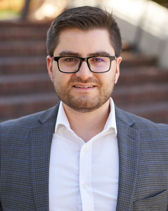
Zaid Atto, Founder and CEO of Xpan Inc.

“Both funds helped us attract new capital to the company and allowed us to advance from a proof-of-concept stage to a functional technology ready for regulatory submission,” Atto says. “Support and backing from FACIT also helped enhance our credibility in the wider ecosystem, as it does for many other start-ups.”

This commitment to supporting oncology innovation and the entrepreneurs behind it underpins all FACIT’s initiatives. Backed by deep private sector expertise, FACIT is uniquely positioned to invest substantive and patient capital in very early-stage innovations. This approach has led to exciting results – to date, FACIT’s investments have helped launch 18 Ontario start-ups and attract an additional $1.4 billion to the province.

“Our unique commercialization-venture model has all the key components to advance innovations at a critical point along their development pathway. Fostering Ontario-First commercialization will ultimately lead to greater impacts for the province’s economy and the health of its people,” O’Neill says.

In Biswas’s experience, FACIT’s engagement with Ontario innovators is unrivaled in the province and is helping meet an unmet need. “The level of support provided by FACIT is truly unique in Ontario’s innovation ecosystem,” Biswas says. “My team and I are deeply thankful for FACIT’s ongoing commitment to empowering local start-ups like Tenomix to grow roots and scale in Ontario.”

OICR works with its strategic partner FACIT to ensure made-in-Ontario breakthrough technologies have a clear path from the lab to patients. For more about how OICR and FACIT work together visit https://oicr.on.ca/about/commercialization-and-facit/

Making liver cancer ablations more accurate with 3D ultrasound guidance

An OICR Imaging project has developed a 3D ultrasound system that provides real-time feedback on the best placement of thermal ablation applicators.


To fully destroy a liver tumour, Dr. Derek Cool must guide his thermal ablation applicator needle into a patient’s abdomen, avoiding vital organs and blood vessels, to the exact centre point of a tumour.

“If I’m off centre by just a couple of millimetres, the ablation might not cover the entire tumour and the patient won’t get a complete treatment,” says Cool, an Interventional Radiologist at London Health Sciences Centre and Assistant Professor in Western University’s Department of Medical Imaging.  

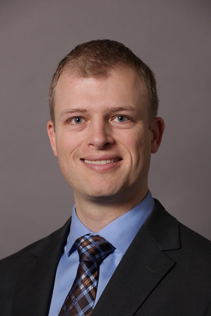
Dr. Derek Cool

Thermal ablation is a procedure where heat is used to kill cancer tissue. It’s highly effective, less invasive than traditional surgery and used around the world, but outcomes vary because of the challenge of placing the applicator needle so precisely. With such small margins for error, the procedure takes coordination and dexterity from the physician and a clear view of the tumour, the needle and the surrounding tissue.

That view is usually provided by one of two imaging tools ⁠— ultrasound or CT scan ⁠— but both have their limitations. Ultrasound delivers real-time images, but only on a 2D plane. CT scans provide a more wholesome view, but not in real-time. And access to CT is limited, especially in smaller healthcare centres and in the developing world.

Cool wanted to create a more accessible tool to make thermal ablations for liver cancer more accurate, so he teamed up with OICR’s Director of Imaging Dr. Aaron Fenster to develop a 3D ultrasound guidance system and test it in an ongoing clinical trial.

The system they developed is compatible with standard ultrasound equipment, making it a potential low-cost upgrade for healthcare organizations. It works by attaching a 2D ultrasound transducer to a robotic arm that moves it around and takes images at different angles. Those 2D images are synthesized by a computer program that generates a 3D image.

“Ideally, our system provides a 3D view of the whole tumour and allows the procedure to be done entirely with ultrasound, without having to occupy a CT scanner,” Fenster says.

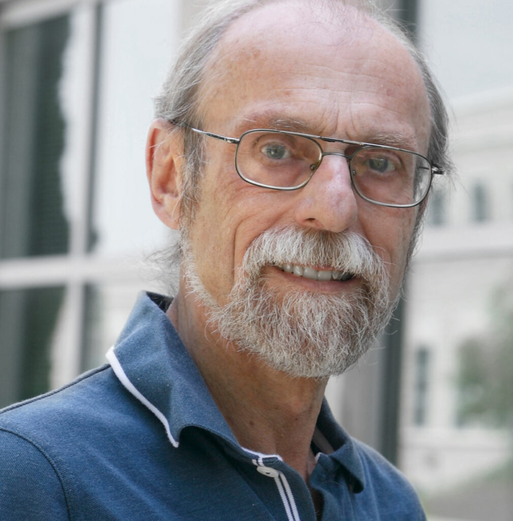
Dr. Aaron Fenster

The first half of their trial focused on feasibility. The 20 ablation procedures performed by Cool and his interventional radiology colleagues were guided by 2D ultrasound, and 3D images were collected to be reviewed afterward. From these first 20 patients, they found that it was easy to incorporate the 3D ultrasound system into the ablation procedure and the 3D images effectively showed the needle position within the tumour.

The 3D ultrasound data from these early patients showed that the needle insertion with 2D US was accurate, yet improvements in the needle position was still possible. They worked with colleagues to develop software that uses 3D ultrasound data to analyze the position of the applicator needle, predict how likely a full ablation is from that position, and provide guidance on where to move the applicator for a more complete ablation. In a recent IEEE Transactions on Medical Imaging paper, they reported that feedback from their software could have improved needle placement in eight of 14 procedures.

“The physician needs to know when they start the ablation if it will be successful,” Fenster says. “Our software tells them whether the needle is too deep or too shallow, or needs to move up, down, left or right in order to be successful.”

In the next phase of their trial, Cool aims to use 3D ultrasound as part of the procedure to confirm needle positioning and suggest adjustments, which is how he thinks it could be most helpful as part of a radiologist’s workflow. Though he has performed more than 200 successful ablations, he says he often finds himself wanting another angle to pinpoint the centre of the tumour.

“The 3D ultrasound is going to improve our ability to assess where our applicators are and confirm if we’re going to get full tumour coverage,” he says.

3D ultrasound systems developed in Fenster’s laboratory have been patented and licensed to multiple companies. With rates of liver cancer rising in Ontario and around the world, Cool and Fenster hope their system can help make thermal ablation more accessible, especially when expensive CT scanners are not accessible.

“We hope to make this procedure much more accurate and less variable, and potentially change the paradigm of how patients are treated in Ontario and around the world,” Fenster says.

Susan Fitzpatrick named Chair of OICR’s Board of Directors

OICR welcomes Ms. Susan Fitzpatrick as the new Chair of the OICR Board of Directors. Fitzpatrick, who has been on OICR’s Board since March, brings extensive experience in Ontario’s healthcare sector to the role, as well as strong track record of organizational leadership. She is currently Head of the Canadian Drug Agency Transition Office at Health Canada developing proposals to establish this agency.

Fitzpatrick previously served as interim CEO of Ontario Health, where she oversaw the establishment of the new agency which combined 20 provincial and regional health agencies with $30 billion in funding and more than 12,000 employees. Fitzpatrick also served as CEO of the Toronto Local Health Integration Network and as Associate Deputy Minister in Ontario’s Ministry Health and Long-Term Care.

“We are excited and proud that Susan is our new Chair. Her incredible leadership, knowledge and experience will assist the Institute in improving outcomes for patients and their families by maximizing our impact on Ontario’s healthcare system,” says Dr. Laszlo Radvanyi, President and Scientific Director, OICR. “On behalf of the entire OICR community I welcome her to this role and thank her for her contributions to our work and improving the lives of cancer patients.”

“This is an exciting new opportunity for me to contribute to Ontario’s cancer system with a dynamic organization that is leading cutting-edge research,” says Fitzpatrick.

Read Susan Fitzpatrick’s bio

New prognostic biomarkers and cancer-driving genes found in the dark matter of the cancer genome

Catalogue of 166 biomarkers generated through machine learning analysis of long non-coding RNAs

Toronto – (November 15, 2021) A new batch of prognostic cancer biomarkers have been discovered in an area where few have gone looking before. Researchers have unveiled a catalogue of 166 prognostic biomarkers, generated by analyzing long non-coding RNAs (lncRNAs) – which are understudied in cancer research. Further, one biomarker within the catalogue was shown to be highly effective in categorizing gliomas (brain cancers) as low- or high-risk. The findings demonstrate the potential of lncRNAs as clinical biomarkers and potential therapeutic targets, break new ground in biomarker and cancer biology research and add to emerging science about the role of non-coding RNA dysregulation in cancer.

The study, published in Cell Reports and co-led by Dr. Jüri Reimand, Principal Investigator at the Ontario Institute for Cancer Research (OICR), and Dr. Daniel Schramek, Principal Investigator at the Lunenfeld-Tannenbaum Research Institute (LTRI), used machine learning to evaluate 5,600 potential lncRNA biomarkers against nearly 9,500 cancer samples across 30 cancer types. This narrowed the field to 166 lncRNAs that were found to correlate with patient survival. In a clinical setting these biomarkers could potentially be used to boost the predictive value of clinical variables, molecular features and cancer subtypes to better forecast patient outcomes.

Within their catalogue, the researchers zeroed in on one lncRNA, called HOXA10-AS, that their initial machine learning analysis revealed was a strong candidate as a prognostic biomarker to stratify patients as having either a low- or high-risk brain cancer. To confirm the results of their machine learning analysis, they tested it on a new brain cancer data set, which was generated by co-authors based in Shanghai, China, led by the Huashan Hospital.

Encouraged by the machine learning results, the team moved onto functional biological validation using patient-derived cancer cells, xenograft and organoid models. These steps not only confirmed that HOXA10-AS can act as a robust biomarker, but also as a potential therapeutic target since it plays a role in several important biological pathways in brain cancer. For example, overexpression of HOXA10-AS was associated with increased cell invasion while reduced levels inhibited cell proliferation – both important hallmarks of brain cancer.

These biological experiments also provided further evidence of ‘switch-like’ behaviour exhibited in HOXA10-AS and its link to patient outcomes. Lack of expression of this lncRNA was associated with low-risk brain tumours and high expression associated with aggressive tumours.

“We are excited by the results of this study which not only yielded new biomarkers and insights into cancer biology, but also provides motivation to continue our exploration of the cancer transcriptome for new discoveries to help patients,” says Reimand, whose team led the machine learning analysis. “We have only begun to scratch the surface of the role of RNAs in cancer and are poised for more discoveries as whole-transcriptome sequencing becomes more commonplace in the clinic and more data is available.”

“Functionally annotating cancer genomes and identifying novel biomarkers of patient survival, but also genes that regulate the aggressiveness of cancers, is of uttermost importance for our goal of precision oncology, the idea to improve cancer therapy based on the genetic alterations found within a tumour,” says Schramek, whose team led the functional analysis. “A deep understanding of the factors that drive cancer initiation and progression will facilitate the identification of novel therapeutic targets and treatment strategies.”

“Congratulations to OICR, LTRI and its research partners for making this important new discovery in cancer research,” says Jill Dunlop, Minister of Colleges and Universities. “Ontarians can be proud of the leading role OICR played in this initiative, which could lead to many more cancer discoveries that support better patient outcomes in the future. OICR has an impressive track record of helping to move oncology discoveries and innovations to real world application, and their success is reflected in some of the ground-breaking work taking place at our world-class universities and research institutes.”

“The findings of Dr. Reimand and his collaborators show that there is great value in studying the non-coding regions of the cancer genome. The discovery of robust biomarkers and new therapeutic targets is critical to developing the next generation of precision medicine,” says Dr. Lincoln Stein, Head, Adaptive Oncology, OICR. “I congratulate the entire team on these important findings, which have enriched our understanding of cancer biology and present new opportunities to improve the clinical management of cancer.”

Study puts spotlight on FDA’s accelerated approval of cancer drugs

Ten drugs given accelerated approval by the FDA remained recommended and in-market even if later found not to increase patient survival compared to standard of care

During the HIV epidemic of the 1990s, there were few drugs to combat the virus. With more-promising drugs in testing, pressure grew on the U.S. Food and Drug Administration (FDA) to speed their approval to get them to patients in desperate need. In response, an accelerated approval pathway was developed that allowed new drugs to be approved based on early evidence of benefit. This practice eventually grew to include cancer drugs, which now make up most of the drugs granted accelerated approval. Now, a study recently published in the British Medical Journal has found that in the end, many cancer drugs with accelerated approval have not proven to be of benefit to patients but continue to be used in the clinic.  

“The entire premise of accelerated approval is that a drug is allowed to enter the market under the condition that confirmatory studies are conducted within a reasonable amount of time and that they look at key clinical endpoints like overall survival and quality of life,” explains Dr. Bishal Gyawali, co-author of the study and Associate Professor at Queen’s University and OICR Clinician-Scientist. “However, some studies are delayed by years and often continue to use less-robust surrogate endpoints as evidence of benefit. In addition, even when no improvement in primary clinical endpoints is shown, the FDA can be slow to withdraw its approval and clinical guidelines do not always match the available evidence.”

To grasp the scale of this problem, Gyawali and his collaborators scanned the FDA database for all cancer drugs that had received accelerated approval between the creation of the pathway in 1992 and December 2020. Their analysis found that 10 cancer drugs failed to demonstrate clinical benefit in a combined 18 indications in confirmatory trials, all of which used overall survival as their primary endpoint and compared those drugs against the standard of care. However, a third of these indications for these drugs were allowed to remain on-label for an average of four years. The researchers also found that in most cases, even after a drug’s approval for a given indication had been withdrawn or revoked by the FDA, it continued to be strongly recommended in the National Comprehensive Cancer Network (NCCN) clinical guidelines.

Gyawali theorizes that a number of factors have led to issues with the accelerated approval pathway. He says it is a complex problem but highlights that the FDA faces immense pressure from pharmaceutical companies, patient advocacy groups and physicians to provide wider access to the latest drugs. Conflicts of interest may also be at play, given what Gyawali describes as a “revolving door” between officials at the FDA and the pharmaceutical industry.

“Accelerated approval is a fantastic pathway in theory, but how has it become broken, against the benefit of patients? In many cases it is not fulfilling the compromise between speed and evidence as was intended,” says Gyawali. “It is very understandable that patients want access to the latest drugs, but would patients want these drugs if they knew they were of no benefit? Not acting on the evidence is doing them a great disservice at an immense economic cost, given that in the U.S. Medicare must pay for drugs that are recommended in NCCN guidelines.”

NCCN guidelines are used globally, including in Canada, but Canada’s system of determining drug approval and funding has so far prevented the issues seen with accelerated approval in the U.S. “In Canada we have a two-step process with Health Canada approving drugs and a separate body (CADTH) deciding if they should be publicly funded,” he explains. “There are calls by some to combine the Canadian decision-making pathway into one so that any newly approved drug is automatically funded, but I think we need to treat the U.S. experience as a cautionary tale. Of the 18 indications that we highlight in our paper, most are not publicly funded in Canada. This demonstrates that our current system is working well, although there is always room to improve.”

Another aspect to improving decision-making to select the most effective new agents is robust quality-of-life assessments in clinical trials as well as real-world data through patient-reported outcome measures in addition to the clinical efficacy of new drugs. These factors need to be increasingly balanced into the whole equation, together with ‘toxicities’, both physical and financial.

Gyawali acknowledges that the problems with the FDA’s accelerated approval pathway are complex but says that there are avenues to improve the situation. “Really it all comes down to education. If we can train the next generation of oncologists to think differently about drugs and policy, they can become a force to push for improvements in the system and the type of information that is being used to inform treatment decisions,” he says.

“On the other side of the coin is patient advocacy groups. Some patient groups are also misled to believe that newer is always better. I speak to many patient groups to help them understand the nuances of such regulatory policies when it comes to cancer drugs. We need to make them allies in our efforts to improve the system so that it is of true benefit to patients.”

New AI tool enables analysis of millions of cells within minutes

Method eliminates the need for manual identification of cell types

Tumours are more than just a clump of cancer cells. Within them you will also find stroma cells connecting tissue, various immune cells, and other types. Understanding this cellular makeup is essential to many areas of cancer research, especially biomarker discovery. Recently, a team of Toronto-based researchers unveiled a new AI-based method of analyzing data from imaging mass cytometry (IMC) and other high parameter technologies used to determine a sample’s cellular profile. Their work is described in a recent article in the journal Cell Systems.

“One of the big questions in this field is how do you go from the raw data generated by highly multiplexed imaging such as IMC to actually determining the presence and abundance, or absence, of different cell types,” explains the study’s lead author Dr. Kieran Campbell, Investigator at Lunenfeld-Tanenbaum Research Institute and an OICR Affiliate researcher. “Right now, if you take a tissue section and run it through IMC there is quite a laborious, manual process of analyzing the resulting data that is open to subjectivity due to the need for human annotators to assign cell types.”

IMC provides a high-dimensional view of a tumour sample by mapping the expression of about 40 different proteins. Analysis of the data allows for single cells within a sample to be assigned to a type. To address the shortcomings of current methods of analysis Campbell and his collaborators created “Astir” (ASsignmenT of sIngle-cell pRoteomics) to automate the process of annotating cells, doing away with the need for human annotators and offering other advantages.

“Current methods of automation can only go as far as putting individual cells into groups and then it’s off to the human annotators to determine what the cells in those groups are. However, with Astir we can automate the assignment of cell types for each individual cell within a sample based on previously known biology,” explains Campbell. “Astir is also able to do this at great speed – in our study we showed that the system could assign a cell type to 800,000 cells in 15 minutes on a standard desktop computer.”

Campbell and his collaborators, which include Dr. Hartland Jackson, OICR Investigator, envision two primary uses for this new technology. In a discovery setting, researchers could use Astir to conduct an initial broad analysis of a dataset to get a high-level picture of the cellular makeup of their samples. In addition, with further development, the research group sees future clinical use for their invention to look for predictive or prognostic cell types within a single sample. “This tool is crucial for both our ability to analyze millions of cells from each clinical sample we investigate and within these samples identify rare cells that may play an important role in cancer development or progression,” explains Jackson.

“Infiltration of a tumour by immune cells is a prognostic indicator across many cancers, but to infer this from IMC currently requires a lot of manual analysis and expertise,” explains Campbell. “So this is really the first step towards automating that. We look forward to continuing development to include elements such as the spatial location of the cells within a sample with the goal of enabling new discoveries and ultimately helping patients.”

Clinical trial breaks new ground on using immunotherapy for more cancer types

Guided by blood samples, collaborative research team shows that patients’ tumour genomic and immune landscape predict response to a common immunotherapy

A Phase II clinical trial has shown that regular monitoring of circulating tumour DNA (ctDNA) in patients can be used to predict and assess the efficacy of the immunotherapy pembrolizumab. Building on that previous finding, a new study of the same patients’ tumours, co-led by Drs. Trevor Pugh and Lillian Siu, and recently published in Nature Communications, produced clinically significant findings that have bolstered understanding of how genomic and immune characteristics shape response to this therapy. The study, part of a clinical trial called INSPIRE at the Princess Margaret Cancer Centre (PM), was the first to test pembrolizumab across a range of metastatic cancers such as head and neck, breast and ovarian cancers and rare solid tumours.

“From previous research we knew that the level of ctDNA in the bloodstream is very strongly associated with response to this therapy – increases in ctDNA after treatment are predictive of poor outcomes,” explains Pugh, Director of Genomics and a Senior Investigator at OICR and a Senior Scientist at PM. “By looking at ctDNA and response to this immunotherapy across many different cancer types, we were able to get a better understanding of the underlying mechanisms – both immune and genomic – that determine how well this therapy works for an individual patient.”

The research team’s pan-cancer approach (studying across cancer types) allowed them to discover that in non-responding patients, many different molecular mechanisms were at play. “It was quite striking to see that many of these tumours had their own unique way of evading immune response from the therapy,” says Pugh. “It shows the need to break out of the cancer type-specific mold and expand our investigations to include the immune cells that infiltrate tumours and play a role in patient outcomes.”

These important findings were the result of a successful multi-disciplinary and inter-institutional collaboration that brought together researchers and clinicians from several organizations and specialties. The OICR-PM Joint Genomics Program and the PM Tumour Immune Profiling and Cancer Genomics Programs, with support from industry partners Natera and Merck, worked together to discover the new biomarkers.

“This study is a perfect example of what you can accomplish by taking a team approach to science. We are indebted to the study team, our patients and their families for their support.” says Siu, a Senior Scientist and medical oncologist at PM and the BMO Chair in Precision Cancer Genomics. “Through INSPIRE, not only did we discover new biomarkers, we also laid the groundwork for new investigator-initiated trials so that we can continue to find even more ways predict response to immunotherapy.”

It is anticipated that the knowledge generated from this study will be incorporated into clinical genomics reports generated by the OICR-PM Joint Genomics Program by screening patients for biomarkers of response to pembrolizumab and other immunotherapies so that the patients most likely to benefit enroll in these trials.

“As someone who leads a clinical lab, this really excites me,” says Pugh. “By building this new knowledge into the workflow of our CAP-accredited lab we can provide patients with another potential treatment option and help enable this type of therapy for more types of cancer, as it has only been commonly used in lung cancer and melanoma, with notable success.”

The study found that there are some shared ‘immunogenomic’ traits that predict response to pembrolizumab. For example, women with breast and ovarian cancers generally had a poor response to the therapy, but those that did respond shared molecular characteristics with melanoma patients who had similar outcomes.

“The work of our multidisciplinary team is allowing us to explore new uses of immunotherapies and will help us deliver even more useful information to patients and their doctors,” says Pugh. “We are excited to continue our work on this less-invasive method of managing patient care and are planning more studies to enable increased use of ctDNA and genome sequencing in the clinic.”

Read more: Princess Margaret Cancer Centre researchers expand understanding of immunotherapy benefits (UHN newsroom)

This work was supported by the Princess Margaret Cancer Foundation, Ontario Institute for Cancer Research, Terry Fox Research Institute, Gattuso-Slaight Personalized Cancer Fund, BMO Chair in Precision Cancer Genomics, Canada Research Chair in Translational Genomics, Canada Foundation for Innovation, Ontario Ministry of Research and Innovation, and Merck for the study drug.