Hands-on lab internship reaffirms research aspirations for Sudbury student

OICR sponsored fourth-year undergrad Chelsea Leduc to study in a molecular biology lab through the 2025 BioCanRx Indigenous Summer Student Internship.

It was during the summer after Chelsea Leduc’s first year of university at the University of Ottawa that she discovered her interest in science went beyond math and physics.

She spent that summer volunteering at a cancer centre in her hometown of Sudbury. Inspired by the cancer patients she met, she decided to switch programs from chemical engineering to biomedical science.

“Being around patients and understanding their experiences really motivated me to want to pursue a career in medical science,” Leduc says.

Now, a few years later, another summer experience has shown Leduc she is on the right track.

OICR sponsored Leduc for the 2025 BioCanRx Indigenous Summer Student Internship, a collaboration between OICR, BioCanRx and the Canadian Cancer Society that provides Indigenous students with hands-on research or policy experience.

Leduc interned in the lab of Dr. Sujeenthar Tharmalingam at the Northern Ontario School of Medicine, where she contributed to molecular biology research about radiation resistance in triple-negative breast cancer.

“This summer confirmed how much I really enjoy research, and reaffirmed this is the path I want to go down,” says Leduc, who is now entering her fourth year of undergraduate studies.

OICR News asked Leduc about her experiences during her internship and her plans for the future.


How was your experience in Dr. Tharmalingam’s lab?

It was a great experience. My supervisor was very helpful and supportive — we talked about my goals and tailored my experience to work toward them.

During the internship, I got the opportunity to study the mechanisms that underly resistance to radiation in triple negative breast cancer, and in particular to validate genes that had previously been identified as potentially related to radiation resistance. Through this and other work, I was able to contribute to an academic paper in progress, and get a lot of valuable experience.

What were the most interesting aspects of cancer research you learned about during your internship?

Some of the most interesting things for me were fundamental skills. For example, I really enjoyed cell culturing. You have your own set of cells to take care of, and you need to come in every single day to check on them. It made me realize that research is a lot more than coming into the lab and doing an experiment. I also did a lot of qPCR testing. I don’t even know how many plates I ran this summer, but I got pretty proficient at doing them successfully.

These are both such important skills, and they will my transition into other labs in the future much easier.


How did the internship shape your perspective on your future career path?

This internship helped me see the importance of research and how it can translate into improved to patient care, and that reinforced my interest in research as well as my interest in medicine. It also helped boost my confidence and gave me a clearer sense of direction in my career path. I’m excited by the possibilities in front of me, including a master’s degree, a PhD and even medical school.

Why are internship opportunities like this important for students?

I think they’re incredibly valuable because they give students like me the opportunity to get a taste of what research looks like outside of an undergrad lab. A career in research is a big commitment, and it’s really important to be able to experience what it’s like before making a decision on your future.

National guideline for Lynch Syndrome aims to prevent cancers and save lives

Dr. Raymond Kim co-led the development of 18 recommendations to improve outcomes for Lynch Syndrome, a condition that increases the risk of multiple cancers.

Experts in cancer, genetics and medicine came together with patient partners to publish evidence-based guidelines for managing Lynch Syndrome they believe can save lives across Canada.

Co-led by Ontario Hereditary Cancer Research Network (OHCRN) Head Dr. Raymond Kim, the Canadian Lynch Syndrome Working Group set out to improve testing and management of Lynch Syndrome, an inherited genetic mutation in the body’s mismatch repair (MMR) system that increases a person’s risk of developing cancer.

While Lynch Syndrome is the leading inherited cause of colorectal and endometrial cancers, it is not as widely understood as other cancer-causing genes like BRCA1 and BRCA2. This lack of awareness contributes to inconsistent practices around when to test for Lynch Syndrome, and what actions to take once Lynch Syndrome has been diagnosed.

The Canadian Lynch Syndrome Working Group consisted of 37 experts, including geneticists, genetic counsellors, oncologists and patient representatives. After reviewing evidence and conducting a clinical survey, they came to consensus on 18 wide-ranging recommendations that were published in the Journal of Medical Genetics.

Key recommendations include universal Lynch Syndrome screening for people with colorectal and endometrial cancers, genetic testing for family members of people with Lynch Syndrome, and the creation of provincial surveillance protocols for Lynch Syndrome-associated cancers.

“These recommendations could improve outcomes for people with Lynch Syndrome by finding cancer earlier or even preventing it altogether,” says Kim, a Clinician Scientist and Medical Geneticist at University Health Network’s Princess Margaret Cancer Centre, Sinai Health System and The Hospital for Sick Children.

Another of the group’s recommendations is for all Canadian jurisdictions to create a provincial/territorial registry of hereditary cancers, which is exactly what Kim has spearheaded with OHCRN.

OHCRN was created by OICR to build a centralized database on hereditary cancers in Ontario that can drive new discoveries to detect, diagnose and treat hereditary cancers. The Network’s participant portal will officially launch in November 2025.

Diagnosing and managing hereditary cancers have been a key focus for OICR in recent years. In 2023, Kim and OICR’s Dr. Trevor Pugh were part of a team that developed a blood test that can detect cancer in people with Li Fraumeni Syndrome — another cancer-causing genetic condition. Kim and Pugh are also part of the CHARM Consortium, which is exploring similar tests for people with Lynch Syndrome. Also in 2023, Kim and OICR’s Dr. Harriet Feilotter contributed to guidelines for hereditary cancer screening that were adopted into practice by Ontario Health.

For the new Lynch Syndrome guideline to be adopted into practice, Kim says the recommendations will need to be endorsed by provincial agencies and new infrastructure will need to be built. But he says implementing the recommendations would make a huge difference for people with Lynch Syndrome.

“This is an important opportunity to standardize care, improve equitable access and save lives,” Kim says.

Cancer Research Changed My Life: Jerry’s story

Dr. Jerry Battista describes what it was like to receive treatment for prostate cancer using techniques he helped develop.

I was a researcher in cancer on the side of medical physics, and for many years worked at trying to improve the precision of radiation therapy treatments. 

It was very strange when I was diagnosed with prostate cancer and I would be receiving radiation as treatment with techniques that I helped develop.

I was diagnosed through a PSA test. The PSA values were going up, and it was time to decide on a course of treatment. I opted for a very compressed schedule of radiation treatments.

This is a major advance resulting from cancer research. The previous protocol would have patients treated over a month or more, and here the radiation treatment is compressed into a week and a half.

It has gone very well. The PSA is under control, there were minor side effects about a year or so after treatment, but they’ve resolved.

In time, I had an opportunity to become a patient partner with the OICR. And it was very tantalizing for me because of my dual role as researcher and then as a patient

Cancer research certainly has changed my life. I am an almost full-time musician now still enjoying performing, and that’s a very nice outcome for me.


Dr. Jerry Battista is a retired medical physicist with expertise in radiation oncology and a survivor of prostate cancer. As an OICR patient partner, he assists researchers who are developing advanced 3D medical imaging.

Technological first in DNA sequencing leads to real-time brain cancer diagnosis

OICR’s Dr. Jared Simpson developed a world-first application for Oxford Nanopore sequencing that is making a difference for patients with brain cancer.

When Dr. Jared Simpson developed the first-ever software to detect DNA methylation using the Oxford Nanopore Technologies sequencing platform in 2017, he knew there was huge potential for cancer research.

Measuring DNA methylation — a process associated with cancer where a cell’s function is changed due chemical ‘tagging’ — had already proven helpful in classifying different types of cancers.

And Nanopore sequencing — an affordable, portable and faster alternative to traditional genome sequencing platforms — had widely recognized potential to make to sequencing easier and more accessible.

But what Simpson couldn’t have known in 2017 was that his software would help launch a series of advancements that has culminated in a UK hospital now using Nanopore sequencing to classify brain tumour types in real time in the operating room.

“We knew our method was state-of-the-art, and that it would be useful for cancer research and hopefully for cancer patients,” says Simpson, Scientific Director and Senior Principal Investigator of Computational Biology at OICR. “But what it has become, with this application to classify brain cancer in a clinical setting, is absolutely amazing.”

Nanopore sequencing is a relatively new technology. It reads larger chunks of DNA at one time and more quickly than traditional “short read” sequencers do. While the per-run cost of Nanopore sequencing is slightly higher than short-read sequencers, the machinery itself is significantly less expensive.

By 2017, Nanopore technology had advanced to the point where it could process enough data to sequence a human genome, while remaining small enough to sit on a desktop. So, the time was right for Simpson and colleagues to apply this new sequencing technology to DNA methylation.

“We knew it was possible, but we had to figure out how to train machine learning models,” Simpson recalls. “Importantly, we also wrote software that anyone could use to predict methylation from Oxford Nanopore sequencing data.”

They published their work in a 2017 Nature Methods paper and made their software package available with open access. In the years since, their software was used by several research teams studying DNA methylation and became widely recognized as the gold standard in the field.

The software was of particular interest to researchers studying brain cancer, which has dozens of molecular subtypes that can be diagnosed using methylation and are critical to determining the best ways to treat a tumour. The speed and accuracy of Simpson’s software on the Nanopore sequencing platform allowed researchers to explore quicker, more effective tools to diagnose brain cancer subtypes that could ultimately be used to guide treatment.

Simpson’s software laid the groundwork for further advancements to Nanopore sequencing methods, including a 2023 Nature study that used a newer deep-learning based method to detect methylation for ultra-fast classification of brain tumour types.  

Then early in 2025, a team led by Dr. Matthew Loose of the University of Nottingham (UK) took a major step forward in the use of Nanopore to classify brain tumours. Loose and colleagues’ advanced software allows all necessary brain tumour subtyping tests to be combined into one assay performed on Nanopore, cutting the time it takes to accurately diagnose brain tumours from a few weeks to a few hours.

The process is so quick that, during trials at a Nottingham hospital, some tumours were sampled and sequenced within the same surgery, allowing doctors to make surgical decisions based on the results.

“What we are seeing is that clinicians obtain results faster, patients can move to the appropriate treatment and care pathways faster and patient uncertainty is reduced,” Loose says.

Dr. Matthew Loose

This groundbreaking clinical application of Nanopore has the potential to transform how brain cancers are diagnosed, providing patients with quicker results that can be actioned sooner and give them the best chance at surviving cancer. It builds on years of work, much of which was enabled by Simpson’s 2017 innovation.

“Jared’s early work…was foundational to the translational applications we are now developing with Nanopore sequencing,” Loose says.

For Simpson, whose lab is largely focused on developing computational methods and software to understand cancer, it’s gratifying to see his work making a difference in the clinic.

“This direct clinical application to classify people’s brain cancer within the operation room — that’s very exciting and confirmation that what we do is having an impact on patients,” Simpson says.

As sequencing technology continuing to advance, Simpson sees even more exciting clinical applications on the horizon. His lab is now using Nanopore sequencing to find cancer DNA in blood samples — tests that are often referred to as ‘liquid biopsies’, and have been shown to detect cancer earlier than imaging and other existing tests.

“Nanopore is cheap, portable and increasingly more accurate,” Simpson says, “and so it could help bring important genomic testing to hospitals and healthcare centres across Ontario.”

Cancer Research Changed My Life: Iain’s story

Iain Bancarz explains how a career in bioinformatics at OICR has changed his life and given him the opportunity to help others.

I started my career in computer science. With a PhD in computer science, I had many career options. 

I decided to get into bioinformatics because it was taking off shortly after I finished my PhD. It was very exciting — lots of opportunities, lots of great work being done. 

It was good, but it was a little bit abstracted. It was a little bit separate from immediate applications. 

So I began thinking about cancer research. Also, I wanted to get into this field for more personal reasons. 

Like many people, I have a personal connection to cancer. My uncle passed away about 10 years ago. He was quite young — he was only in his 50s and he left behind three children in their 20s. So he got to see his kids grow up, but his grandkids have never got to meet him. 

I can identify with that because I never met one of my own grandparents because he too passed away from cancer.

Working in cancer research is an opportunity to give something back, to help people see their grandkids grow up or to meet their grandparents when they otherwise might not have been able to. 

This career change involved relocation for me because I’d spent a lot of my career in the UK. The reason I moved specifically to Toronto is because of the Ontario Institute for Cancer Research because I’d heard about it from colleagues. I’d heard it was a good place and it was doing good work. 

It really did change my life. And here I am now. I love living in this city and doing the work that I do. And I really am happy and proud to be with such great colleagues because everyone here knows that we are working to help people with cancer.


Dr. Iain Bancarz is a computational biologist and manager of the Clinical Genome Interpretation team at OICR. He is originally from Edmonton, lived much of his life in the UK, returned to Canada in 2018 and has worked for OICR since then. He loves cycling and once did a sponsored bike ride from Cambridge, England to Paris, France to raise funds for cancer research.

AI-generated genomes could accelerate precision medicine without compromising patient confidentiality

OncoGAN generates simulated genomes that can be used to train genomic analysis tools without the confidentiality concerns associated with real genomes.

A new AI system that creates simulated cancer genomes could reshape the tools used to analyze tumours, helping bring about more accurate cancer diagnosis and ultimately more effective treatments.

OncoGAN was developed by researchers at the Ontario Institute for Cancer Research (OICR) and the University of Toronto and is described in a new Cell Genomics paper.

It uses generative AI to simulate realistic tumour genomes across eight different types of cancer, including breast, prostate and pancreatic cancers. These synthetic genomes can simulate realistic patterns of genetic alterations, and can be used to benchmark genomic testing and improve the algorithms that make ‘precision oncology’ possible.

Analyzing tumour genomes and the variations within their DNA has enabled new discoveries about how cancer develops, leading to a surge of cutting-edge tests and medicines. It is the cornerstone of precision oncology, where cancer treatment is personalized to the unique biology of a patient’s tumour.

But the algorithms used to analyze genomes are limited because they have been trained on a limited set of cancer genomes, relatively few of which are publicly available. The most commonly used tools were trained on a few dozen legacy genomes, and can’t fully capture the necessary biological diversity. While more recent genome sequencing data exists, access is often restricted due to concerns around the confidentiality of the patients they were sampled from.

“With OncoGAN, we are creating realistic genomes out of nothing, with no connection to any real person, yet they have a huge amount of value scientifically,” says Dr. Lincoln Stein, Scientific Director (Acting) at OICR, Professor of Molecular Genetics at the University of Toronto, and senior author of the paper. “These synthetic genomes don’t contain any personal health information, and so they can be shared without limitation.”

Beyond privacy, another advantage of OncoGAN’s synthetic genomes is that their exact ‘ground truth’ is known. A genome’s ground truth is its full, error-free DNA sequence with all genomic variants identified. It is nearly impossible to know the ground truth of real-life genomes because they are so complex and sequencing technology is limited. This means that current genome analysis tools could be flawed, because there may have been trained on flawed data.

Dr. Ander Díaz-Navarro

By generating genomes from scratch, OncoGAN gives researchers fully known, verified DNA sequences that can enable better, more precise genomic testing and analysis.

“Knowing the ‘ground truth’ of the genomes means they can be used to benchmark new algorithms with full knowledge of that the correct answer is,” says Dr. Ander Díaz-Navarro, Postdoctoral Fellow at OICR and first author of the paper.

With better, more accurately trained tools to analyze cancer genomes, Stein says scientists could unlock more critical insights with the potential to transform cancer care.

“The more we know about the biological factors that drive cancer, the better equipped we are to detect it as early as possible, treat it more effectively, and even prevent it altogether,” Stein says.

OncoGAN is publicly available for download. Stein, Díaz-Navarro and colleagues have also generated 800 simulated genomes, which are available with open access and are already being used to train analysis tools in Stein’s lab.

Study reveals young-onset breast cancer risk for women taking hormone therapy

A global collaboration highlights the need for personalized approaches for treating various conditions involving fluctuating hormone levels.

An international study published in The Lancet Oncology is the largest and most comprehensive to highlight the links between hormone therapy and the risk of breast cancer in women under the age of 55.

Researchers pulled together data from 13 cohort studies across North America, Europe, Asia and Australia — including The Canadian Study of Diet, Lifestyle and Health, co-led by OICR’s Dr. Victoria Kirsh — as part of a large collaboration known as the Premenopausal Breast Cancer Collaborative Group. They found one type of hormone therapy increased women’s risk of early-onset breast cancer, while another may reduce their risk.

Hormone therapy is prescribed to manage symptoms related to menopause or following gynecological surgeries like the removal of the uterus (hysterectomy) or the ovaries (oophorectomy), as well as other conditions affecting hormones levels.

Dr. Victoria Kirsh

Previous research had found links between hormone therapy and breast cancer risk in women above 55 — who more likely to be taking hormone therapy. But premenopausal women also face symptoms caused by fluctuating hormone levels.

“Symptoms necessitating hormone therapy aren’t confined to post-menopausal women,” says Kirsh, who is Interim Director of the OICR-hosted Ontario Health Study. “They can happen to peri-menopausal and younger women, who may then consider hormone therapy, so it’s important they understand the risks involved.”

Between the 13 cohorts, the study analyzed data from 459,476 women ages 16 to 54, about 15 per cent of whom reported having used hormone therapy.

They found dramatically different risks of breast cancers depending on the type of hormone therapy taken. Women who took a combination of estrogen and progesterone saw their risk of young-onset breast cancer jump by 18 per cent, while women who took estrogen alone saw their risk of young-onset breast cancer reduced by 14 per cent.

The risk of young-onset breast cancer among women taking estrogen and progesterone was particularly high for triple-negative disease compare to other subtypes.

Kirsh says the results largely mirror the risks for women over 55.

“The takeaway here is that we need personalized approaches to menopausal symptom management,” Kirsh says. “When women consider taking hormones, particularly combinations of estrogen and progesterone (which is necessary for women with an intact uterus to counteract the increased risk of endometrial cancer associated with estrogen-only therapy), they need to weigh the benefits of symptom relief against the associated risks of breast cancer.”

New PanCuRx Co-Lead aims to bring latest pancreatic cancer innovations to patients

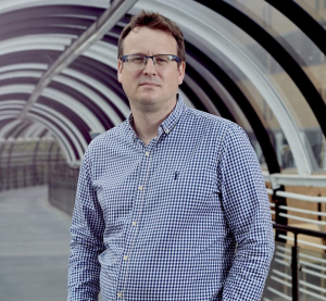
Clinician Scientist Dr. Robert Grant will help lead OICR’s signature pancreatic cancer program into next phase.

Dr. Robert Grant recently became Clinical Co-Lead of OICR’s cutting-edge pancreatic cancer research program, PanCuRx, following the retirement of Dr. Steven Gallinger. As a Clinician Investigator at Princess Margaret Cancer Centre (PM), Grant treats patients with pancreatic and biliary tract cancers and conducts research into integrating Big Data and AI technologies into clinical decision making.

Grant works out of the Wallace McCain Centre for Pancreatic Cancer at PM, which collaborates extensively with the PanCuRx program on clinical trials. He joined OICR News for a chat in which he explained his journey from studying economics to testing drugs on patient-cell derived organoids.

Can you tell us about how you became interested in medicine and research and pancreatic cancer in particular?

When I was completing my undergraduate degree in economics and trying to figure out what I wanted to do, I had been volunteering at the cancer centre in London, and it was a really informative experience. It was inspiring to see everyone there working to help people with cancer, and in addition, my grandfather had cancer at the time. I didn’t know much about oncology, but I decided to pivot my studies to try and get into medical school. To get the necessary prerequisites, I pursued my MA in economics and was successfully accepted to medical school at the University of Toronto.

During my first month there, I was connected with Dr. Steven Gallinger. I heard he was doing cool research, in particular exome sequencing, which was a brand new technology. I remember doing a PubMed search for ‘exome’ and there only being 20 or so papers. It was super interesting to me and Steve’s passion for pancreatic cancer research really rubbed off on me. It was the right place, the right time, I liked the new technology that nobody really understood, and I had some statistical background to bring to the table. I really never looked back from there. 

How does that background in economics play into your work?

I think in many ways, when I was studying economics, it was at the forefront of Big Data and statistical analysis. A lot of the innovative techniques were coming from the economics world which I found interesting, and now Big Data and its analysis are part of fields like medicine and genomics. Economics provided me with a solid foundation for applying these techniques to pancreatic cancer. In addition, I think there’s a broader way of thinking in economics that has probably stayed with me.

Can you tell us about your work as a clinician-investigator? 

As a medical oncologist at the Princess Margaret Cancer Centre, I mostly see people with pancreatic and biliary cancers, many of whom enrol in our innovative clinical trials testing new therapies and other technologies. I also oversee the pancreatic cancer genetics and screening program. Preventing pancreatic cancer remains a central challenge for us, but today, genetics is high impact, since when we find a genetic cause of a pancreatic cancer, we’re able to prevent other cancers throughout a whole family. 

This clinical work blends nicely with what our team at PanCuRx does from a research perspective and my personal lab, which is focused primarily on applications of AI and machine learning in the clinic. These applications include integrating different data types such as electronic health records, wearables and genomic data to improve decision making when it comes to treatment and supportive care. For example, in a recent paper in the Journal of Clinical Oncology, we showed how an AI could help get palliative care to those who need it most. In the end, the goal is to improve outcomes and quality of life.

What are the barriers to getting these tools into the clinic?  

I think all the ingredients are there, but we haven’t made the meal yet, if that makes sense. The algorithms we have are extraordinarily capable and there’s no doubt in my mind that if applied appropriately to the right situation, that they’d have a major impact. However, data is always an issue. We need to have appropriate safeguards and privacy, but we also need a way to let people who want to contribute their data to AI research do so. By sharing their data, people are helping make these models even more useful. We are making progress in this area, but there is still a lot of work to do.

Also, there is still the question of integrating them into routine clinical practice. We need to map out the complex process of providing pancreatic cancer care and think about the people involved in it. Can we embed these tools into existing practices, or do we need to update our practices more fundamentally? How are we going to measure their impact? Some of these technologies, such as reasoning language models, are poised to have broad impacts on care, so these are the complicated questions we need to be asking. I think that these issues represent the ‘last mile’ to clinical adoption, but I am happy to say that there is a lot of good work going on in this area.

What’s it like being part of PanCuRx and OICR?

I’m really grateful to be a part of this community. OICR has been integral to the success we’ve had so far in our pancreatic cancer research, owing in large part to the Institute being a world-leading genomics powerhouse, thanks to the efforts of Dr. Trevor Pugh and his group, working closely over many years with the co-lead of PanCuRx, Dr. Faiyaz Notta. The technologies available at OICR powered our first wave of discoveries and the new technologies being developed here are allowing us to build upon them and continue to innovate.

In terms of PanCuRx, I am proud of the program’s achievements so far and excited about the foundation they provide our group with going forward. We had the COMPASS trial which proved that you can indeed get rapid high-quality genomic data on a metastatic pancreatic cancer using technologies like whole-genome and transcriptome sequencing and laser capture microdissection, and that this data can improve care. We also have the on-going Prosper-PANC trial which is enabling us to evaluate this approach across Ontario.

Building on the COMPASS trial, we recently completed the PASS-01 trial, led by Dr. Jennifer Knox at the McCain Centre for Pancreatic Cancer with Dr. Elizabeth Jaffee from John Hopkins and Dr. David Tuveson from Cold Spring Harbor. This was an international study that evaluated the two standard forms of chemotherapy for pancreatic cancer in a randomized clinical trial. The results of this trial are allowing us to use biology to help in making the key clinical decision of who should get what treatment. OICR’s sequencing expertise and the other scientific specialties provided by our collaborators really allowed us to dig into this question like never before.

Is there anything going in the program right now that excites you in particular?

It’s the fact that we have really branched out across the spectrum of disease. We are working on tests for early detection, have trials in the surgical and radiotherapy spaces and there continues to be a deep focus on biomarkers to help guide treatment in all of our research. The NeoPancONE trial, which was presented at the American Society of Clinical Oncology conference last month and showed that chemo before surgery is beneficial in some subtypes of pancreatic cancer, is a great example of this.

From a more personal perspective, I am really excited about one trial I am leading from a clinical perspective, called ADOPT. In this trial, we are taking cells from a given patient’s tumour and growing living models of that tumour, called organoids, outside of the body. We will then be able to test many different drugs on them and see which ones the patient’s cancer is sensitive to, including drugs we may not normally use. The science is being led by Faiyaz and his team as part of PanCuRx. We hope that this approach can provide patients with treatment options that they didn’t have before, and on top of that, it will be treatments uniquely suited to their case.

More generally, I see a great momentum in the program and in pancreatic cancer research in general and I am really excited to be a part of it. We have built an amazing team, world-class resources, and capabilities spanning from the bench to the bedside, coordinated by the McCain Centre at PM and PanCuRx at OICR. Together with the incredible opportunities more broadly at OICR, with the network of brilliant people and cutting-edge new technologies, I believe we can use pancreatic cancer as a “launch pad” at OICR and PM, bringing the most exciting innovations rapidly into clinical care to make an impact for this devastating disease.

Using AI to interpret prostate cancer MRIs could reduce radiologist workload and give more patients access

A machine learning tool was able to accurately triage prostate cancer in OICR-supported research.

New research suggests that AI has the potential to improve the efficiency of prostate cancer screening, while also raising important questions about the use of AI in healthcare.

In an OICR-supported study published in Abdominal Radiology, Dr. Masoom Haider and his PhD student Emerson Grabke used a machine learning algorithm to interpret the results of mutiparametric MRI (mpMRI) and simulated its potential use as a triaging tool. They found it could reduce radiologist workload with minimal loss in accuracy.

It’s widely recognized that triaging suspected prostate cancer with mpMRI can spare patients and the health system from unnecessary biopsies. That’s why Cancer Care Ontario recommends all patients at risk of clinically significant prostate cancer get an MRI first, before determining if a biopsy is necessary.

But the increased use of MRI for triaging prostate cancer has put extra pressure on radiologists to interpret the results, which led researchers to wonder if AI could help reduce the added burden.

To answer this question, Haider’s lab trained a model on more than 2800 MRI exams. The AI model used a U-Net based convolutional neural network and combined this with clinical risk factors. The model was then run in a simulation on over 460 patient exams to see what would happen if only cases the deemed concerning were reviewed by the radiologist.

Emerson Paul Grabke

“We looked at how many exams could be triaged by U-Net combined with clinical indicators without needing a radiologist’s report, and how many instances of cancer might be missed with this triaging,” says Emerson Paul Grabke, PhD Candidate in Haider’s lab and first author of the paper.

They found that combining U-Net reading the MRI with prostate cancer risk indicators was able reduce radiologists’ workload by about 12.5 per cent with only a small reduction in the number of missed cancers (3 per cent) and a significant reduction in overcalls by radiologists.

“There is some promise here to potentially reduce healthcare costs and deal with shortages in radiologists, while helping more patients access MRI,” says Haider, Director of the Machine Learning and Radiomics Lab at Sinai Health and Professor of Medical Imaging at the University of Toronto.

But Haider says the study was meant to push the envelope into areas that would likely not be acceptable in the current healthcare environment. It raises important questions about our comfort level relying on AI to make important healthcare decisions.

Artificial intelligence (AI) has been touted for its potential to help with clinical decision making, but when and how AI should be used in place of human expertise has been the topic of much debate with human oversight deemed essential.

“We need to have a discussion in the medical community about what level of performance would make this kind of use of AI acceptable — further work is needed,” Haider says.

Haider says these conversations have become even more important as the healthcare system continues to face unprecedented demand. In June, the Ontario government announced it was adding 35 new centres for MRI and CT scans to keep up with increased pressures, which will ultimately means more need for radiologists to review the scans.

“There are interesting opportunities for AI to improve access in situations where it’s harder to get radiologist review — whether that’s late at night in the emergency room or in a remote area of the country,” Haider says. “So, it’s important we get this right.”

New OICR Investigator brings a dynamic approach to uncover the origins of cancer

Dr. Federico Gaiti uses a suite of advanced techniques to study how cancer begins and progresses.

One of OICR’s newest Investigators is bringing together a powerful combination of cutting-edge tools to better understand how cancer develops and spreads.

Dr. Federico Gaiti integrates genetic, epigenetic and spatial genomics approaches with novel computational frameworks to study cancer at the single-cell level, hoping to unlock new ways to diagnose and treat it.

“We combine different types of data to study the changes that occur in cells as they transition toward cancer — not just identifying what those changes are, but also understanding why and how they happen,” says Gaiti, a Scientist at the Princess Margaret Cancer Centre (University Health Network) and Assistant Professor at the University of Toronto.  

Gaiti’s scientific journey began in Italy, where he was born and raised. After earning his bachelor’s and master’s degrees in biology in his home country, he moved to Australia for his PhD studies, followed by postdoctoral research in New York.

As his research evolved from studying non-model organisms during his PhD to focusing on cancer during his postdoctoral training, Gaiti became increasingly interested in the molecular mechanisms driving cancer development. This growing focus led him to Toronto, drawn by its world-class cancer research ecosystem. In 2021, he moved to Canada and established his lab at the Princess Margaret Cancer Centre.

Now, three-and-a-half years later, his lab has grown to a team of eight with diverse skill sets, and they have recently celebrated a major milestone: the publication of their first research paper fully conceptualized and executed in Toronto, featured in Developmental Cell.

OICR News spoke with Dr. Gaiti about the new paper, his lab’s broader research goals, and how becoming an OICR Investigator is helping propel his work forward.

In simplest terms, how would you describe your research focus?

We start with a few fundamental questions: How does cancer begin? How does it become more aggressive over time? And how does it develop resistance to treatment? To answer these, we study the changes that occur in individual cells — not only genetic mutations in the DNA (known as somatic mutations), but also non-genetic changes that influence how cells grow, divide, and interact with their environment.

We use cutting-edge tools and integrate multiple layers of data — including genetic, epigenetic, and spatial information — all at the single-cell level. This comprehensive approach allows us to trace the molecular changes that drive cancer development, with the goal of understanding when and how a normal cell becomes cancerous.

What sort of impact do you hope this work has on patients?

By understanding these changes at the cellular and molecular level, we aim to uncover vulnerabilities in the process of cancer development that could be targeted by new, more effective therapies. Gaining insight into these processes at their earliest stages may also lead to the development of diagnostic tests that detect cancer sooner, when it is most treatable.

Your lab just published its first paper. What did you find in that study?

Our study focused on glioblastoma, one of the most aggressive forms of brain cancer. We discovered that invasive glioblastoma cells ‘hijack’ normal brain development programs, behaving like immature brain cells. We believe this helps explain how these cancer cells spread so effectively throughout the brain. Understanding this process could open new ways to target glioblastoma, not just by trying to kill the cancer cells, but by disrupting the developmental pathways they co-opt to invade healthy tissue.

How has becoming an OICR Investigator helped you realize the goals of your research?

Becoming an OICR Investigator helps bring our ideas to life by connecting us with a network of outstanding scientists and providing access to valuable resources and support. This kind of collaboration is essential for driving science forward. As an early-career researcher, I also deeply value the mentorship opportunities, being able to learn from and seek guidance from more experienced investigators is incredibly helpful in navigating challenges and advancing our work.