
CanPath adds Saskatchewan cohort, all 10 provinces now represented

This post was edited and republished with the permission of CanPath.

CanPath is pleased to announce that, with funding support from the Canadian Partnership Against Cancer, a Saskatchewan cohort will be developed and join the CanPath study. The Saskatchewan Partnership for Tomorrow’s Health (Saskatchewan PATH) will add approximately 9,000 participants to the existing cohort of over 330,000 Canadian participants. The addition of Saskatchewan means that all 10 Canadian provinces have now joined CanPath.

Saskatchewan PATH will create a platform and resource for fostering research in cancer and chronic disease prevention within the province. The Saskatchewan PATH study will be led by Scientific Director, Riaz Alvi and hosted by the Saskatchewan Cancer Agency.

“We are excited to officially welcome Mr. Alvi and the Saskatchewan PATH team to the CanPath partnership. We look forward to working together to develop a truly pan-Canadian study and sharing learnings from our other regional cohorts to support Saskatchewan PATH as they move forward,” says John McLaughlin, Executive Director of CanPath.

 “We are proud to be a part of this truly national program.  Saskatchewan holds a prominent place in the history of healthcare in Canada, and houses one of the world’s oldest cancer registries.  We are confident that the people of Saskatchewan will welcome this opportunity to participate in Saskatchewan PATH to help further a better understanding of cancer and other chronic diseases, and to assist with the future development of prevention, early detection, diagnosis and treatment programs.  There is exciting and highly rewarding work ahead of us.” says Riaz Alvi, Scientific Director of Saskatchewan PATH.

Saskatchewan has a unique and diverse population, with roughly half living in the province’s largest city, Saskatoon, or the provincial capital of Regina. The province’s economy is primarily associated with agriculture and more recently mining. The burden of cancer in Saskatchewan is significant with about 5,600 new cancers diagnosed in 2018 and just over 2,000 cancer deaths in the same year. In 2018, the number of people living with cancer that had been diagnosed within the last 5 years (5-year prevalence), was approximately 17,000 people.

“Since CanPath began almost 11 years ago, we have sought to ensure representation of all provinces. Now being able to include participants from the province of Saskatchewan fills an important gap, and builds upon the hard work of many of us who started and have maintained the CanPath cohort and vision since the beginning,” says Philip Awadalla, National Scientific Director for CanPath.

With CanPath’s guidance and support of the development of Saskatchewan PATH, the new cohort will benefit from the experience and lessons learned by CanPath’s other regional cohorts. Saskatchewan PATH joins the six regional cohorts that currently makeup CanPath: BC Generations Project, Alberta’s Tomorrow Project, Manitoba Tomorrow Project, Ontario Health Study, CARTaGENE (Quebec), and Atlantic PATH.

The development of Saskatchewan PATH will consist of three phases:

  • Phase I – Planning & Implementation (Present to March 2022)
  • Phase II – Participant Recruitment and Collection of Data and Biological Samples
  • Phase III – Maintenance and Use of Participant Data and Biological Samples

About CanPath

The Canadian Partnership for Tomorrow’s Health (CanPath) is Canada’s largest population health cohort and a national platform for health research. Comprised of more than 330,000 volunteer participants, CanPath is a unique platform that allows scientists to explore how genetics, environment, lifestyle and behaviour interact and contribute to the development of cancer and other chronic diseases. CanPath is hosted by the University of Toronto’s Dalla Lana School of Public Health with national funding from the Canadian Partnership Against Cancer. The Ontario Institute for Cancer Research (OICR) hosts CanPath data in a safe and secure environment. To learn more, visit www.canpath.ca.

The original post can be viewed here: https://canpath.ca/2020/09/canpath-completes-provincial-map-with-addition-of-a-saskatchewan-cohort/
Deploying liquid biopsies to improve cancer screening and care during COVID-19

OICR-supported researcher Dr. Harriet Feilotter leads liquid biopsy research program

Adapted from Canexia Health’s news release and Patriot One Technologies’ news release.

As the COVID-19 pandemic has impacted many areas of life, including the diagnosis and treatment of other health conditions, people have chosen to forgo cancer screening and care in attempt to minimize their potential exposure to the virus. Relative to the general population, people living with cancer are more susceptible to the virus, but delaying cancer treatment may allow the disease to grow or spread.

Dr. Harriet Feilotter has teamed up with members of the pan-Canadian Digital Technology Supercluster to bring greater access to cancer testing and treatment during the pandemic and beyond. Through the $2.59 million Project ACTT (Access to Cancer Testing & Treatment in Response to COVID-19), they aim to provide liquid biopsy solutions, which require only a simple blood draw, as alternatives to surgical tissue biopsies for cancer diagnosis and care.

“The goal is to allow patients alternatives to invasive procedures that may be difficult to access during a pandemic,” says Feilotter, Molecular Geneticist and Scientist at Kingston Health Sciences Centre, faculty member of Queen’s Cancer Research Institute and OICR Associate. “Not only would this benefit those patients who live far from large cancer centres, but it could limit patient exposure to COVID-19 and increase health system capacity.”

The collaborative team is led in part by Canexia Health, which develops specialized cancer genomic assays, and Patriot One Technologies Inc.’s subsidiary Xtract AI, which specializes in machine learning solutions across a variety of applications, among other private and public partners. Together, they will work to enhance their current tests that detect mutations in circulating tumour DNA (ctDNA) from blood and deploy these tests for multiple cancer types across Canada.

Now through ACTT, some patients have access to these tests in British Columbia, Ontario, Quebec and Saskatchewan. The long-term objective is to increase access across the country.

“The development of liquid biopsies and ctDNA testing has been accelerated by this pandemic,” says Feilotter. “We’re proud to team up in this cross-disciplinary, cross-sector collaboration to bring these promising solutions to more patients.”

Read Canexia Health’s news release or Patriot One Technologies’ news release

Analyzing SARS-CoV-2: A cancer researcher trainee’s perspective

OICR-based PhD Candidate awarded University of Toronto COVID-19 Student Engagement Award

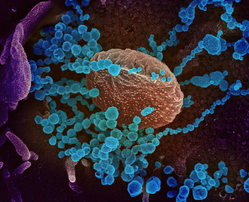
This scanning electron microscope image shows SARS-CoV-2 (round blue objects) emerging from the surface of cells cultured in the lab. SARS-CoV-2, also known as 2019-nCoV, is the virus that causes COVID-19. The virus shown was isolated from a patient in the U.S. Credit: NIAID-RML

When the COVID-19 pandemic shut down labs across Canada, cancer research trainees looked for ways to help respond to the pandemic. PhD candidates Tom Ouellette and Jim Shaw saw an opportunity to combine their skills and contribute to the cause.

Ouellette and Shaw were recently awarded a University of Toronto COVID-19 Student Engagement Award for their project titled Network and evolutionary analysis of SARS-CoV-2: A vaccine perspective. Together, they will develop new machine learning tools to analyze the SARS-CoV-2 genome and how it evolves. 

Tom Ouellette, PhD Candidate in Dr. Philip Awadalla’s lab at OICR.

“We’re two like-minded individuals with complementary skillsets who enjoy coding, math and solving problems, which – fortunately – can be done remotely,” says Ouellette, who is a PhD Candidate in Dr. Philip Awadalla’s lab at OICR. “We saw the opportunity to help with COVID-19 research and we’re happy to apply our skills to help advance research towards new solutions for this pressing problem.”

Ouellette specializes in evolution and population genetics and Shaw specializes in network analysis and algorithm development. Through this award, they will investigate how SARS-CoV-2 is evolving by looking into specific regions of the virus’ genetic code from samples around the world, using mathematical modelling, machine learning, and evolutionary simulations. They are specifically interested in how these changes in the genetic code may alter the virulence, or severity, of the virus.

Jim Shaw, PhD Candidate in mathematics at the University of Toronto.

“Just like cancer, different pressures or stresses can make viruses evolve,” says Shaw, who is a PhD Candidate in mathematics at the University of Toronto. “Understanding these changes can have an impact on how we build vaccines. Furthermore, better understanding of the virus’ evolution may shed light on viral reinfection, which is an important issue as we move into the later stages of the pandemic.”

Ouellette and Shaw plan to publicly release the code that they develop through this initiative for other researchers to build upon.

“SARS-CoV-2 has a much simpler genome than a cancer genome, so it can serve as a simplified model to test out new analytical techniques,” says Ouellette. “Ultimately, I hope to bring the tools and technology we create back into my research on cancer so we can better understand how cancer evolves and becomes resistant to treatment.”

Read more on how OICR researchers are helping understand and overcome COVID-19

Understanding how cancer differs between sexes: A deeper dive

Connie Li
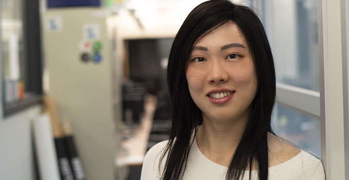
Constance Li, PhD student and researcher in OICR’s Computational Biology program.

In the most comprehensive analysis of whole cancer genomes to date, OICR researchers identify novel sex-linked genomic differences that may be able to predict cancer severity and response to therapy

Cancer differs in males and females but the origins and mechanisms of these differences remain unresolved. A better understanding of sex-linked differences in cancer could lead to more accurate tests and allow sex to be included as a consideration when personalizing treatments for patients.

In a study, published in Nature Communications, OICR’s Constance Li and collaborators identify key genetic characteristics that differ between sexes. Here, Li describes what they found and what this means for patients.

Some studies have already hinted that cancer genomes differ between males and females. What is new about this study?

Previous studies focused on the exomes of patient tumours. That means that they were only looking at a small fraction of the genome that codes for proteins. This study allowed us to look at the entire genome – all of our DNA code – and take a dive deep into many aspects of the disease, like how tumours evolve over time.

By looking at the entire genome and in this ‘dark space’ that we hadn’t explored, we were able to confirm some previous findings but also find new differences between male and female tumour samples.

What sort of differences did you find?

We catalogued the differences we found across nearly 2,000 patient tumours representing more than two dozen different cancer types. Interestingly, we found that biliary cancers – like some liver, gall bladder and bile duct cancers – evolve differently in males than they do in females.

We also found that mutations in the TERT promoter – which is a hot topic in cancer research – occur much more often in men than in women, especially in thyroid cancers.

What does this mean for researchers who are looking into this subject?

Our findings suggest that there are underlying biological differences in the way that male and female tumours begin and progress. Overall, we need to be aware of these differences and consider the sex differences as we develop new tools that can match patients to appropriate treatments.

How else could this be helpful for cancer patients?

These findings are preliminary but powerful. It is important to note that more clinical data and research are needed to validate the differences we found. Ultimately, if we look deeper and find that a cancer progresses along one course in females and a different course in males, we can design roadblocks – or therapies – to stop the cancer along that specific course for that sex.

This paper is part of the Pan-Cancer Analysis of Whole Genomes Project. Read more about the Pan-Cancer project here.

New findings pave the way for future brain cancer research and drug development

The future of brain cancer research

OICR-supported study finds key mechanisms driving a severe form of brain cancer affecting infants and toddlers

When a young child is diagnosed with ependymoma, their treatment options are limited to surgery and radiation therapy – the latter of which causes severe side effects to the developing brain. Despite several clinical trials, scientists have yet to identify life-extending chemotherapies for this type of brain cancer.

In an OICR-supported study recently published in Cell, a research team at The Hospital for Sick Children (SickKids) re-examined how scientists have been studying ependymoma and invented new ways to model the disease. Their work has uncovered key mechanisms behind these tumours and new approaches to treat them.

Lead authors Dr. Antony Michealraj and Sachin Kumar, who are both members of Dr. Michael Taylor’s lab, discussed these promising findings with OICR News.

What spurred this research question?

Dr. Antony Michealraj.

AM: Unfortunately, treatment options for young children with ependymoma are very limited. Radiation treatments led to severe side effects and the disease often returns, so we are very motivated to develop new therapies for these infants and toddlers.

Our previous research showed that these brain tumours emerge very early in a child’s development and, remarkably, there are no specific genetic mutations that are known to cause these tumours. Instead, these tumours possess a unique way of regulating what genes are on or off – a unique epigenetic profile.

We observed that patient tumours have an enriched hypoxia (oxygen level) signature which is correlated with poor survival. These unusual scenarios pushed us to study how hypoxia and epigenetics are linked in ependymoma to search for potential solutions.

How did you approach this challenge and what did you find?

AM: The first problem that we faced was the availability of relevant disease models. What we realized was that we could not study the disease unless it was in a very specific environment with fine-tuned oxygen levels. In the body, these cancer cells only grow in low oxygen and we needed to mimic such an environment. Once we did so, we ended up with an exceptional experimental model of ependymoma that nobody has been able to create before.

These models allowed us to study the microenvironment of ependymoma cells. We saw that the cellular metabolism, or how a cell consumes and uses nutrients, was responsible for the epigenetic dysregulation seen in patients. Using an array of metabolic and epigenetic inhibitors, targeting these pathways destroyed ependymomas, providing an avenue for novel therapeutic interventions.  

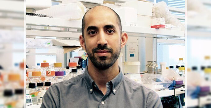
Sachin Kumar.

SK: One exciting finding was what we call our “Goldilocks” model. The key was histone lysine methylation – a process regulating how DNA is wrapped and coiled in a cell. Ependymoma cells require a very fine balance of histone lysine methylation, and too much or too little results in the cells dying.

By studying how to keep these cells alive, we learned how we could potentially eliminate them. The idea would be to find or repurpose drugs that target these pathways within the body, creating an unfavorable environment and eliminating them for good.

How can we translate these discoveries into new therapies for patients?

SK: With our new knowledge of the key molecular pathways involved in ependymoma, we can now look to develop specific compounds – or potential drugs – that can alter these pathways, disrupt the cancer cell’s environment, and prevent these tumours from growing. These compounds may include drugs that are already in clinical studies or completely new molecules. What’s great is that now we have a model that we can use to screen these drugs more effectively.

AM: We can screen FDA-approved drug libraries on these disease models which will enable us find potential chemotherapies rapidly. Since there are currently no approved medicines that work for this type of brain cancer, if we find a drug that works, it could potentially become the standard of care for this disease around the world.

We hope that these findings pave the way for future therapy development. Although we’re in the very early stages of developing any new drugs, we understand how important this work is to the children and families affected by the disease. We’re committed to finding new solutions for them.

Read more about our achievements in brain cancer research on OICR News.

Q&A with new OICR investigator Dr. Hartland Jackson on the latest in mass cytometry, single-cell imaging and his return to Canada

OICR welcomes Dr. Hartland Jackson back to Toronto as Lunenfeld-Tanenbaum Research Institute and OICR’s newest investigator

While he was a doctoral student developing experimental models of breast cancer, Dr. Hartland Jackson recognized the enormous potential impact of multiplexed imaging and single-cell technologies. If we could see how different cells interact within a tumour, what could we discover?

This question fueled his research over the last half decade, taking him to Switzerland to develop advanced imaging methods alongside experts at the University of Zurich. Now, returning to Canada, Dr. Jackson plans to collaborate across disciplines and sectors to apply this technology to solve more scientific and clinical questions. Here, he discusses his goal of bringing the benefits of this technology to more patients in Ontario and around the world.

What was your main research focus in Switzerland?

HJ: In a nutshell, I was developing a new technology, called imaging mass cytometry, which allows us to visualize and analyze tumour samples in more detail than ever before.

When I joined the research group at the University of Zurich, they had developed a prototype imaging system. My role was to take this system and be the first to apply it to a clinical problem. Ultimately, I helped shepherd the system from a prototype to a commercial product that is now used around the world.

What clinical application did you focus on?

HJ: I focused on investigating how this technology could help in the diagnosis and prognosis of breast cancer. Through this process, we made a lot of progress in developing analysis methods and optimizing the system. Whereas traditional imaging methods could see three or four markers on a cell, our system allows us to see 40 markers at the same time. With this technology and imaging system, we could visualize how different cells were organized within a sample, which revealed new types of breast cancer.

In addition to this discovery, my work showed that imaging mass cytometry can reveal information within clinical samples – meaning information that may be useful for patients. We pushed the boundary on what can be done with this system and now it’s used around the world to study different human diseases.

Interestingly, the technology that I was working on was an adaptation of an earlier technology developed in Toronto by DVS Sciences, which was supported in part by OICR. My plan was to work with the imaging experts in Switzerland and bring these developments back to the place where the technology was created and is now manufactured as a commercial product by Fluidigm.

Is that what brought you back to Canada?

HJ: Yes, one of the reasons I’ve returned to Canada is to bring this expertise back to Toronto. In addition to that, the research community here is very impressive. The universities, research institutes and hospitals are all tightly knit. This makes for an excellent environment to develop new technologies that can address clinical health challenges. I find that researchers here are like-minded in their goals and collaborative spirit. We enjoy working through technical challenges and delving into the mysteries of cell biology, and – at the same time – working on research that really matters to patients.

What will your future research focus on?

HJ: I plan to continue developing some of the methods that I was working on in Europe while expanding my research in a few exciting areas.

We’re looking to apply this technology to different types of cancer and different diseases in collaboration with clinician scientists. I’m interested in applying this technology in drug clinical trials to help us understand how patients respond to different therapies. In parallel, I look forward to using this technology to study experimental model systems to better understand how cells are communicating with each other and what goes wrong in the communication between cells during cancer development.

Our work has shown what this technology is able to do and that has only opened more avenues for future research. I’m excited because these new applications are now within our reach. To date, collaborations have allowed me to make more progress than I could have ever made on my own and I look forward to building new collaborations to make new discoveries in the future.

Visit Dr. Hartland Jackson’s OICR website page

Mapping the roots of brain cancer

Dr. Hayden Selvadurai, a lead researcher on the project to identify the origins of childhood brain cancer.

OICR-funded researchers pinpoint short-lived cells that give rise to childhood brain tumours

Childhood brain tumours are remarkably complex, but understanding their origins could help researchers develop drugs to eliminate them. Where can these cells be found? How early do they appear? How do they lead to tumours? For Dr. Hayden Selvadurai, these unresolved questions were a call to action.

In a recent study, published in Cell Reports, Selvadurai and collaborators at The Hospital for Sick Children (SickKids) discovered a rare type of stem cell that gives rise to medulloblastoma, the most common type of brain cancer in children. Their study shows that these cells arise early in brain development and exist for a brief period of time – a developmental window which scientists can now home in on.

“If we can’t eliminate the stem cells at the root of medulloblastoma, we can’t effectively treat the disease,” says Selvadurai, who was a Postdoctoral Fellow under the supervision of Dr. Peter Dirks while leading this study. Dirks is Head of the Division of Neurosurgery at SickKids, Principal Investigator at The Arthur and Sonia Labatt Brain Tumour Research Centre, Professor at the University of Toronto and Co-leader of OICR’s Brain Cancer Translational Research Initiative (TRI). “These problematic cells arise amid a complex and intricate process of fetal brain development and we were able to pinpoint exactly when that happens.”

The study builds on the research group’s previous publication in Cancer Cell that traced the origins of medulloblastoma growth back to a small group of cells that distinctively expressed the SOX2 gene. Using single-cell RNA sequencing, lineage tracing and advanced imaging techniques, the team showed that these stem cells were responsible for generating all other tumour cells and could give rise to new tumours if not fully eliminated.

“I’m proud of these findings because we were able to unify our knowledge of developmental neurobiology with cancer biology,” says Selvadurai. “We were able to build on our understanding of medulloblastoma growth while improving our experimental models of brain cancer. Together, this work could help the community develop new effective treatments for patients with the disease.”

Dirks’ research group plans to further investigate the genes involved in the early stages of medulloblastoma in collaboration with OICR’s Brain Cancer TRI team.

This study was supported in part by the Canadian Institutes of Health Research and OICR through the Stand Up to Cancer (SU2C) Canada Cancer Stem Cell Dream Team.

Q&A with Monique Albert: Ontario’s international leader in biobanking

The Ontario Tumour Bank’s longstanding leader appointed Secretary of International Society for Biological and Environmental Repositories

The International Society for Biological and Environmental Repositories (ISBER) today announced the appointment of Monique Albert as Secretary of the Society’s Board of Directors.

With two decades of experience in research and biobanking and three years of experience on the Society’s Board as Director-at-Large Americas, Albert has been re-elected to the Board into the executive role of Secretary.

In her new position, Albert will lead the maintenance of ISBER by-laws, policies and procedures affecting nearly 1,000 ISBER members who lead hundreds of biobanks around the world. While assuming this role, Albert will continue to serve as Director of the Ontario Tumour Bank at OICR, a position that she has held for more than seven years.

Here, she reflects on her new role and her experiences to date.

How did you become involved in preserving human specimens for research?

MA: I began working directly with human specimens as a researcher in 2001, using cutting-edge technologies to analyze human samples. It was through this experience that I realized the utmost importance of preserving and maintaining the quality of these specimens to generate the most reproducible data. Good biological science is built on good data, which can only come from well-preserved samples.

When I recognized the importance of these invaluable samples, I began developing initiatives to improve biobanking practices at my local research institute. I’ve been building on those initiatives ever since.

Quality is an important aspect of your work. How do you make quality maintenance sustainable?

MA: While sample quality is a key element of a biobank’s success, it is not the only one that matters. To be successful, a biobank needs to meet current and future research needs, comply with standards and regulations, and operate in a sustainable way for future generations. I’m fortunate to have a background in project management and business planning that helps balance these three elements with limited resources.

As biobanking has become more mainstream, I’m proud that Ontario has consistently been at the forefront of biobanking standards. I’ve had the privilege of sharing my work with the growing international biobanking community through presenting at conferences and publishing on several occasions.

What are you looking forward to in your new role as Secretary?

MA: Having plenty of experience with ISBER – and ISBER’s savvy, inclusive and collaborative members – I know we are making an incredible impact on research. I’m honoured to be elected to this role and to continue to volunteer my time for the continued growth of ISBER. My previous experience at ISBER will allow me to hit the ground running and keep the momentum on existing goals and initiatives with the best interests of the Society and its members at heart.

Read more about ISBER’s 2020 Election Results or more on Monique Albert’s active role within ISBER on OICR News.  

Study points to common protein duo as a new therapeutic target for several cancer types

Structure of the RUVBL1 protein. (Credit: Emw / CC BY-SA (https://creativecommons.org/licenses/by-sa/3.0))

Local research group discovers a new way to shut down a pair of cancer-driving proteins, pontin and reptin, using the structure of an FDA-approved drug

Pontin and reptin are proteins that are involved in several cancer-driving mechanisms and play key roles in several diseases, including liver, colorectal, breast, lung and bladder cancers. This makes them a hot target for cancer drug development and discovery efforts. Currently, there is only one drug class that may hold some promise to shut down these proteins, but a Toronto-based team of scientists has recently broken new ground.

Dr. Walid Houry’s Lab at the University of Toronto and OICR’s Drug Discovery group have discovered that pontin and reptin, also known as RUVBL1 and RUVBL2, may be blocked to prevent cancer growth using a chemical similar to the FDA-approved drug, sorafenib. Their findings, which were recently published in Biomolecules, could be a starting point for new and improved cancer drugs based on the approved drug’s structure and function.

Dr. Nardin Nano.

“Through our research, we detangled a large, complex process of interactions between proteins, but what we found was both rewarding and exciting,” says first author Dr. Nardin Nano, who was a PhD student in the Houry Lab while leading the study. “Our findings suggest a new target for cancer treatment and that a new therapy could be within reach.”

This study is part of a larger initiative, led by Nano and members of the Houry Lab, to further describe the function of these proteins in helping cancers grow and invade tissues. With their newfound understanding, the Houry Lab will continue to design and develop molecules similar to sorafenib that can better target pontin and reptin.

“I look forward to future studies that will use this knowledge to better inhibit these proteins in vivo,” says Nano. “Although there is more work to be done, I’m proud that this discovery can help guide future drug development efforts.”

“Given the multiple roles of pontin and reptin in carcinogenesis, it’s not surprising that they are promising drug targets,” says Houry, who is a Professor at the University of Toronto and supported by OICR’s Cancer Therapeutics Innovation Pipeline. “These findings motivate us to continue developing pontin and reptin inhibitors as potential anti-cancer compounds that could – one day – help a number of patients with the disease.”

Unexpected mitochondria activity in leukemia cells gives opening for new treatments

Toronto researchers unravel key cancer-driving circuit between the “powerhouse” and the “brain” of leukemia cells, in big first step for future therapeutic discovery and development

Dr. Dilshad Khan.

Over the last few decades, research has suggested that mitochondria, also known as the “powerhouses of the cell”, play an important role in tumour growth and development, but little is known about how to prevent these cellular machines from wreaking havoc. In a recent study, scientists have discovered a key protein that is made in the “powerhouse of the cell”, unexpectedly affects the expression of genes in the nucleus, or the “brain”, of certain leukemia cells. The study was launched by Dr. Dilshad Khan, who – alongside colleagues in Dr. Aaron Schimmer’s lab at the Princess Margaret Cancer Centre – set out to determine which genes in the mitochondria were essential to the growth and viability of acute myeloid leukemia (AML).

Through genome-wide CRISPR screening and other gene-manipulating techniques, they discovered a key mitochondrial protein that AML cells can’t survive without – MTCH2. Their findings, which were recently published in Blood, may eventually lead to new ways to fight this common and fast-growing form of blood cancer.

“We found that the mitochondrial protein MTCH2 is essential for the growth and survival of AML cells,” says Khan, Postdoctoral Fellow in the Schimmer Lab, who is the first author of the study. “But finding this protein was just one piece of the puzzle. We needed to understand how it worked.”

With Khan’s expertise in epigenetics, the team systematically dissected how MTCH2 affects AML cells. They found that blocking this protein would ultimately cause leukemic stem cells – the difficult-to-treat renewable cells that are thought to be at the root of leukemia – to irreversibly transform into cells that are easier to eliminate with existing chemotherapies.

“Through a series of experiments, we unraveled how MTCH2 affects AML cells and discovered that this protein has a remarkable and unexpected impact on nuclear pathways – it could control nuclear gene expression to affect AML stemness and survival,” says Khan. “We never thought this could happen, but now that we’ve discovered these new links, we could potentially find new ways to control these mechanisms.”

Next, the Schimmer Lab and collaborators plan to investigate MTCH2’s specific mechanism to find where inhibitors – or potential cancer drugs – could block its path. These initiatives will add to Schimmer’s research on dysregulated mitochondrial pathways in leukemia, including his recent work on fat production and copper distribution in leukemic stem cells. This research is funded in part by OICR’s Acute Leukemia Translational Research Initiative and OICR’s Cancer Therapeutics Innovation Pipeline.

“This study showed us that mitochondrial proteins are more interconnected with other cellular networks than we thought,” says Khan. “These fundamental findings have shed light on new research avenues that we can pursue to find new solutions that will hopefully benefit patients with AML.”