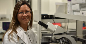
Dr. Bayani’s research focus is on precision medicine approaches to discover and validate biomarkers using large, well-annotated retrospective clinical and clinical trial cohorts.
Translation of biomarkers and their diagnostic platforms are key to prospective clinical trial design and successful uptake in the diagnostic setting. Critical to understanding the variable outcomes seen in the current standard of care treatment for cancers like breast and prostate is recognizing the impact of genomic heterogeneity and serves as models for discovery and translational initiatives in other cancer types. Specifically Dr. Bayani’s team investigates the impact of heterogeneity at the genomic, transcriptomic, and proteomic levels and their integration with other diagnostic testing modalities such as imaging and digital pathology to build a clinical toolbox that can to improve risk assessment and treatment decision-making. This can significantly reduce the unnecessary overtreatment of some patients, while accelerating it in others.
- PhD, Princess Margaret Cancer Centre/Ontario Cancer Institute, Mt. Sinai Hospital, University of Toronto
- MHSc, Princess Margaret Cancer Centre/Ontario Cancer Institute, University of Toronto
- Interim Co-Director, Diagnostic Development, OICR
- Principal Research Scientist, Diagnostic Development, OICR
- Assistant Professor, Department of Laboratory Medicine and Pathobiology, University of Toronto
- Evaluation of multiple transcriptomic gene risk signatures in male breast cancer. Bayani J, Poncet C, Crozier C, Neven A, Piper T, Cunningham C, Sobol M, Aebi S, Benstead K, Bogler O, Dal Lago L, Fraser J, Hilbers F, Hedenfalk I, Korde L, Linderholm B, Martens J, Middleton L, Murray M, Kelly C, Nilsson C, Nowaczyk M, Peeters S, Peric A, Porter P, Schröder C, Rubio IT, Ruddy KJ, van Asperen C, Van Den Weyngaert D, van Deurzen C, van Leeuwen-Stok E, Vermeij J, Winer E, Giordano SH, Cardoso F, Bartlett JMS. NPJ Breast Cancer. 2021 Jul 26;7(1):98. doi: 10.1038/s41523-021-00301-0.
- Comparative survival analysis of multiparametric tests-when molecular tests disagree-A TEAM Pathology study. Bartlett JMS, Bayani J, Kornaga E, Xu K, Pond GR, Piper T, Mallon E, Yao CQ, Boutros PC, Hasenburg A, Dunn JA, Markopoulos C, Dirix L, Seynaeve C, van de Velde CJH, Stein RC, Rea D. NPJ Breast Cancer. 2021 Jul 8;7(1):90. doi: 10.1038/s41523-021-00297-7.
- Jane Bayani*, Elizabeth N Kornaga*, Cheryl Crozier, Gun Ho Jang, Lauren Bathurst, Irina Kalatskaya, Quang M. Trinh, Cindy Q Yao, Julie Livingstone, Paul C Boutros, Melanie Spears, John D. McPherson, Lincoln D. Stein, Daniel Rea , John M.S. Bartlett. Identification of Distinct Prognostic Groups: Implications for Patient Selection to Targeted Therapies amongst Anti-Endocrine Therapy Resistant Early Breast Cancers. Journal of Clinical Oncology Precision Oncology. 2019 May. DOI: 10.1200/PO.18.00373 JCO Precision Oncology – published online May 10, 2019.
- Molecular stratification of early breast cancer identifies drug targets to drive stratified medicine. Bayani J, Yao CQ, Quintayo MA, Yan F, Haider S, D’Costa A, Brookes CL, van de Velde CJH, Hasenburg A, Kieback DG, Markopoulos C, Dirix L, Seynaeve C, Rea D, Boutros PC, Bartlett JMS. NPJ Breast Cancer. 2017 Feb 15;3:3. doi: 10.1038/s41523-016-0003-5. eCollection 2017.
- Singh SK, Hawkins C, Clarke ID, Squire JA, Bayani J, Hide T, Henkelman RM, Cusimano MD, Dirks PB. Identification of human brain tumour initiating cells. Nature. 2004 Nov 18;432(7015):396-401.
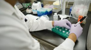
OICR’s Diagnostic Development program focuses on tissue-based analysis with expertise in formalin-fixed, paraffin-embedded (FFPE) human samples and a breadth of associated technologies for complex genome analysis, nucleic acid and protein extraction, tissue microarray construction, NanoString technologies, automated immunohistochemistry and quantitative molecular pathology analysis.
Services available:
- DNA extraction (FFPE or frozen tissue).
- RNA extraction (FFPE or frozen tissue).
- Dual DNA and RNA extraction (FFPE and frozen tissue).
- Histology – Tissue sectioning (FFPE and frozen tissue).
- Histology – Laser capture microdissection.
- Histology – routine H&E stain.
- Low-density tissue microarray construction (50-100 cores).
- High-density tissue microarray construction (200 cores).
- Multiplex qPCR (FFPE compatible).
- Library preparation for targeted sequencing (FFPE compatible).
- FISH (FFPE compatible).
- Quantitative FISH analysis.
- Multiplex CNV analysis (e.g., Multiplex ligation dependent probe amplification -MLPA).
- NanoString-based technologies.
- Automated immunohistochemistry (whole sections and TMA).
- Single or multiple labeling (Chromagenic and FITC).
- Antibody optimization and validation.
- Automated quantitative IHC image analysis.
- Slide scanning and archiving.
To learn how Diagnostic Development can help further your project please contact the team at diagnostic.development@oicr.on.ca.